
Abducens nerve
Citation, DOI, disclosures and article data
At the time the article was created Frank Gaillard had no recorded disclosures.
View Frank Gaillard's current disclosuresAt the time the article was last revised Yuranga Weerakkody had no financial relationships to ineligible companies to disclose.
View Yuranga Weerakkody's current disclosures- Abducens nerve (CN VI)
- Abducens nerve (VI)
- Sixth cranial nerve
- Abducent nerve (CN VI)
- Nervus abducens
- Nervus cranialis VI
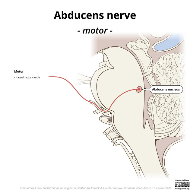
The abducens nerve is the sixth cranial nerve (CN VI). It is a motor nerve responsible for abduction of the eye (TA: nervus abducens or nervus cranialis VI). It courses from the abducens nucleus, located in the dorsal pons, up to the cavernous sinus, via a long cisternal segment that is prone to injury, to its termination on the lateral rectus muscle.
It can be divided into five parts:
- nucleus and intraparenchymal portion
- cisternal portion
- Dorello canal portion
- cavernous sinus portion
- orbital portion
Gross anatomy
Nucleus and intraparenchymal portion
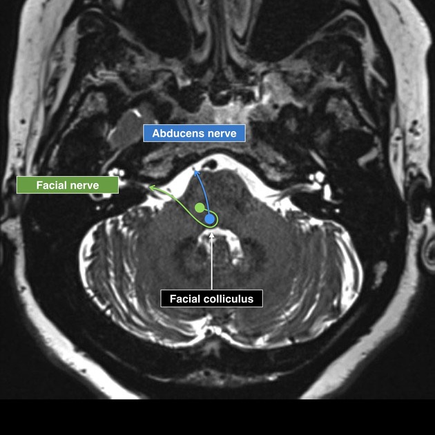
The abducens nucleus is a small nucleus situated at the upper part of the rhomboid fossa beneath the facial colliculus within the pons. Fibers pass anterior through the pons medial to the facial nucleus to reach the pontomedullary junction.
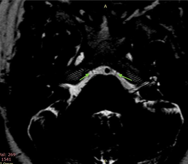
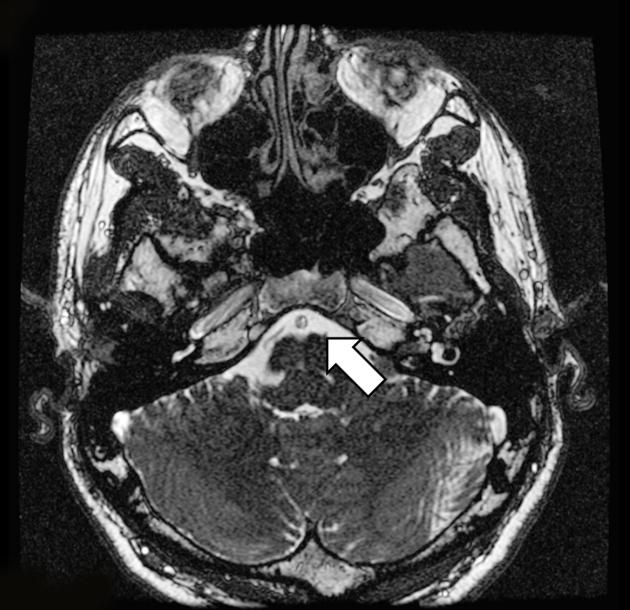
Cisternal portion
The abducens nerve is the most medial of the nerves, emerging immediately below the pons (facial nerve and vestibulocochlear nerve lateral to it) at the pontomedullary junction into the prepontine cistern. It is located within the anterior pontine arachnoid membrane and courses anterosuperiorly towards the petrous apex and cavernous sinus.
Dorello canal
The abducens nerve pierces the dura mater inferior to the posterior clinoid process, enclosed within a fibrous sheath called the Dorello canal, and courses over the medial petrous apex towards the cavernous sinus. Its oblique course and relatively anchored position in the Dorello canal make it prone to stretching when raised intracranial pressure from a space-occupying lesion causes transtentorial herniation (a sixth nerve palsy is the classic lateralizing sign of an extradural hematoma).
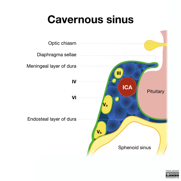
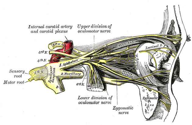
Cavernous sinus portion
Within the cavernous sinus, the abducens nerve is located inferolateral to the internal carotid artery, medial to the lateral wall of the sinus.
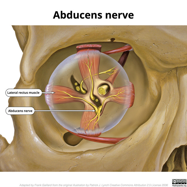
Orbital portion
Having entered the orbit through the superior orbital fissure within the tendinous ring, it supplies the lateral rectus. It is the most inferior nerve passing through the tendinous ring, inferior to the inferior division of the oculomotor nerve.
ADVERTISEMENT: Supporters see fewer/no ads
Related pathology
Quiz questions
References
- 1. Chummy S. Sinnatamby. Last's Anatomy. (2011) ISBN: 9780702033957 - Google Books
- 2. Carmine D. Clemente. Anatomy. (2011) ISBN: 9781582558899 - Google Books
- 3. Umansky F, Elidan J, Valarezo A. Dorello's Canal: A Microanatomical Study. J Neurosurg. 1991;75(2):294-8. doi:10.3171/jns.1991.75.2.0294 - Pubmed
- 4. Kyung Won Chung, Harold M. Chung. Gross Anatomy. (2012) ISBN: 9781605477459 - Google Books
- 5. Sheth S, Branstetter B, Escott E. Appearance of Normal Cranial Nerves on Steady-State Free Precession MR Images. Radiographics. 2009;29(4):1045-55. doi:10.1148/rg.294085743 - Pubmed
- 6. FIPAT. Terminologia Anatomica. 2nd Ed. FIPAT.library.dal.ca. Federative International Programme for Anatomical Terminology, 2019. https://fipat.library.dal.ca/TA2/
- 7. Romano N, Federici M, Castaldi A. Imaging of Cranial Nerves: A Pictorial Overview. Insights Imaging. 2019;10(1):33. doi:10.1186/s13244-019-0719-5 - Pubmed
Incoming Links
- Neurovascular compression syndromes
- Medical abbreviations and acronyms (V)
- Facial colliculus syndrome
- Inferolateral trunk
- Dorello canal
- Basilar artery
- Cavernous sinus haemangioma
- Duane syndrome
- Transition zone (nerve)
- Gasperini syndrome
- Raymond syndrome
- Horizontal gaze palsy with progressive scoliosis
- Superior orbital fissure
- Cavernous sinus contents (mnemonic)
- Superior orbital fissure syndrome
- Pituitary macroadenoma
- Ophthalmoplegia
- Anterior pontine membrane
- Annulus of Zinn contents (mnemonic)
- Cranial nerves
- Trigeminal amyloidoma
- Duane syndrome
- Cranial nerves and brainstem nuclei (illustrations)
- Nerves of the orbit (Gray's illustration)
- Lower pons anatomy - CN VI (diagram)
- Möbius syndrome
- Epidermoid cyst with abducent nerve palsy
- Abducens nerve palsy
- Abducens nerve palsy
- Abducens and facial cranial nerves and nuclei
- Leptomeningeal carcinomatosis
- Normal cranial nerves
- Möbius syndrome
- Abducens nerve on MRI
- Normal cranial nerves
Related articles: Anatomy: Brain
-
brain
- grey matter
- white matter
-
cerebrum
-
cerebral hemisphere (telencephalon)
- cerebral lobes and gyri
- frontal lobe
- parietal lobe
-
occipital lobe
- occipital pole
- lingual gyrus
- fusiform gyrus (Brodmann area 37)
- calcarine (visual) cortex
- cuneus
- temporal lobe
- basal forebrain
- limbic system
- insula
-
cerebral sulci and fissures (A-Z)
- calcarine fissure
- callosal sulcus
- central (Rolandic) sulcus
- cingulate sulcus
- collateral sulcus
- inferior frontal sulcus
- inferior occipital sulcus
- inferior temporal sulcus
- interhemispheric fissure
- intraparietal sulcus
- lateral (Sylvian) sulcus
- lateral occipital sulcus
- marginal sulcus
- occipitotemporal sulcus
- olfactory sulcus
- paracentral sulcus
- paraolfactory sulcus
- parieto-occipital fissure
- posterior parolfactory sulcus
- precentral sulcus
- preoccipital notch
- postcentral sulcus
- rhinal sulcus
- rostral sulcus
- subparietal sulcus
- superior frontal sulcus
- superior occipital sulcus
- superior temporal sulcus
- cortical histology
- cerebral lobes and gyri
- white matter tracts
- deep grey matter
-
pituitary gland
- posterior pituitary and stalk (part of diencephalon)
- anterior pituitary
- inferior hypophyseal arterial circle
- diencephalon
-
cerebral hemisphere (telencephalon)
-
brainstem
- midbrain (mesencephalon)
- pons (part of metencephalon)
- medulla oblongata (myelencephalon)
- white matter
-
grey matter
- non-cranial nerve
-
cranial nerve nuclei
- oculomotor nucleus
- Edinger-Westphal nucleus
- trochlear nucleus
- motor nucleus of CN V
- mesencephalic nucleus of CN V
- main sensory nucleus of CN V
- spinal nucleus of CN V
- abducent nucleus
- facial nucleus
- superior salivatory nucleus
- cochlear nuclei
- vestibular nuclei
- inferior salivatory nucleus
- solitary tract nucleus
- ambiguus nucleus
- dorsal vagal motor nucleus
- hypoglossal nucleus
-
cerebellum (part of metencephalon)
- vermis
- cerebellar hemisphere
- cerebellar peduncles
- cranial meninges (meninx primitiva)
- CSF spaces
-
cranial nerves (mnemonic)
- olfactory nerve (CN I)
- optic nerve (CN II)
- oculomotor nerve (CN III)
- trochlear nerve (CN IV)
- trigeminal nerve (CN V) (mnemonic)
- abducens nerve (CN VI)
- facial nerve (CN VII) (segments mnemonic | branches mnemonic)
-
vestibulocochlear nerve (CN VIII)
- vestibular ganglion (Scarpa's ganglion)
- glossopharyngeal nerve (CN IX)
- vagus nerve (CN X)
- spinal accessory nerve (CN XI)
- hypoglossal nerve (CN XII)
- functional neuroanatomy
- CNS development
- cerebral vascular supply
- arteries
- vascular territories
-
circle of Willis
- internal carotid artery (ICA) (segments)
- vertebral artery
-
normal variants
- intracranial arterial fenestration
- internal carotid artery (ICA)
- anterior cerebral artery (ACA)
- middle cerebral artery (MCA)
- posterior cerebral artery (PCA)
- basilar artery
- persistent carotid-vertebrobasilar artery anastomoses (mnemonic)
- vertebral artery
- ophthalmic artery
-
cerebral venous system
-
dural venous sinuses
- basilar venous plexus
- cavernous sinus (mnemonic)
- clival diploic veins
- inferior petro-occipital vein
- inferior petrosal sinus
- inferior sagittal sinus
- intercavernous sinus
- internal carotid artery venous plexus of Rektorzik
- jugular bulb
- marginal sinus
- occipital sinus
- sigmoid sinus
- sphenoparietal sinus
- straight sinus
- superior petrosal sinus
- superior sagittal sinus
- torcula herophili
- transverse sinus
-
cerebral veins
-
superficial veins of the brain
- superior cerebral veins (superficial cerebral veins)
- inferior cerebral veins
- superficial middle cerebral vein
- superior anastomotic vein (of Trolard)
- inferior anastomotic vein (of Labbe)
-
superficial veins of the brain
-
deep veins of the brain
- great cerebral vein (of Galen)
- venous circle of Trolard
- normal variants
-
dural venous sinuses
- arteries
- glymphatic pathway


 Unable to process the form. Check for errors and try again.