Acetabular labrum
Citation, DOI, disclosures and article data
At the time the article was created Frank Gaillard had no recorded disclosures.
View Frank Gaillard's current disclosuresAt the time the article was last revised Henry Knipe had no recorded disclosures.
View Henry Knipe's current disclosuresAcetabular labrum acts to deepen the acetabulum and increase contact between the pelvis and the femoral head. Its exact biomechanical role remains to be fully elucidated.
On this page:
Gross anatomy
The acetabular labrum is a C-shaped fibrocartilaginous structure with an opening anteroinferiorly at the site of the acetabular notch. Here it is bridged by the transverse ligament (thus forming the acetabular foramen beneath it). Elsewhere it is attached to the margins of the acetabulum.
The labrum is thickest posterosuperiorly and widest anterosuperiorly. It is triangular in cross-section. The fibrocartilage is arranged in three distinct layers:
- external surface: circumferentially oriented layer with radial reinforcing filaments.
- middle layer: dense lamellar collagenous layer
- articular surface: randomly oriented fibrillar layer with chondrocytes
The capsule of the hip joint attaches to the margins or immediately adjacent to the acetabulum and transverse ligament. Superiorly the capsule's attachment is removed from the labrum by a few millimeters forming the perilabral sulcus. Anteriorly and posteriorly the attachment of the capsule is much closer to the base of the labrum, and thus the perilabral sulcus is commensurately smaller.
Variant anatomy
- everted labrum: round or blunted shape on MRI 3
Radiographic features
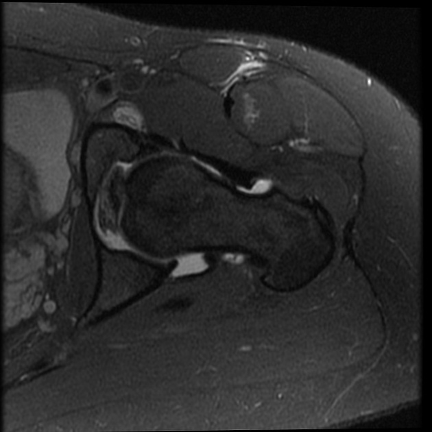
Radiographically, the labrum is best evaluated with MR arthrography and should appear as a uniformly low signal triangular structure, although signal at the base of the labrum is variable. Additionally, intrasubstance heterogeneous signal is more frequently seen in older patients.
Related pathology
Labral pathology contributes to hip pain and the development of osteoarthritis of the hip. Labral lesions are more frequently seen in patients with 'abnormal' hip morphology including:
- cam-type and pincer type femoroacetabular impingement
- acetabular dysplasia
- acetabular labral calcification
References
- 1. Chatha D & Arora R. MR Imaging of the Normal Hip. Magn Reson Imaging Clin N Am. 2005;13(4):605-15. doi:10.1016/j.mric.2005.08.012 - Pubmed
- 2. Petersilge C. Imaging of the Acetabular Labrum. Magn Reson Imaging Clin N Am. 2005;13(4):641-52. doi:10.1016/j.mric.2005.08.015 - Pubmed
- 3. Vogel L, Kraeutler M, Jesse M et al. The Everted Acetabular Labrum: Patho-Anatomy, Magnetic Resonance Imaging and Arthroscopic Findings of a Native Variant. Arthroscopy. 2022;38(1):72-9. doi:10.1016/j.arthro.2021.04.038 - Pubmed
Incoming Links
Related articles: Anatomy: Lower limb
- skeleton of the lower limb
- joints of the lower limb
-
hip joint
- ligaments
- muscles
- additional structures
- hip joint capsule
- zona orbicularis
- iliotibial band
-
hip bursae
- anterior
- iliopsoas bursa (iliopectineal bursa)
- lateral
- subgluteal bursae
- greater trochanteric bursa (subgluteus maximus bursa)
- subgluteus medius bursa
- subgluteus minimus bursa
- gluteofemoral bursa
- subgluteal bursae
- postero-inferior
- anterior
- ossification centers
-
knee joint
- ligaments
- anterior cruciate ligament
- posterior cruciate ligament
- medial collateral ligament
- lateral collateral ligament
- meniscofemoral ligament (mnemonic)
-
posterolateral ligamentous complex
- arcuate ligament
- patellar tendon and quadriceps tendon
- anterolateral ligament
- posterior oblique ligament
- oblique popliteal ligament
- medial patellofemoral ligament
- additional structures
- extensor mechanism of the knee
- groove for the popliteus tendon
- knee bursae
- anterior bursae
- medial bursae
- lateral bursae
- posterior bursae
- knee capsule
- lateral patellar retinaculum
- medial patellar retinaculum
- menisci
- pes anserinus (mnemonic)
- ossification centers
- ligaments
- tibiofibular joints
-
ankle joint
- regional anatomy
- medial ankle
- lateral ankle
- anterior ankle
- ligaments
- medial collateral (deltoid) ligament
- lateral collateral ligament
- additional structures
- ankle bursae
- ossification centers of the ankle
- variants
- regional anatomy
- foot joints
- subtalar joint
- mid-tarsal (Chopart) joint
-
tarsometatarsal (Lisfranc) joint
- ligaments
- intermetatarsal joint
- metatarsophalangeal joint
- interphalangeal joint
- ossification centers
-
hip joint
- spaces of the lower limb
-
muscles of the lower limb
- muscles of the pelvic group
- muscles of the thigh
- muscles of the leg
- anterior compartment of the leg
- posterior compartments of the leg
- lateral compartment of the leg
- muscles of the foot
- dorsal muscles
- plantar muscles
- 1st layer
- 2nd layer
- 3rd layer
- 4th layer
- accessory muscles of the lower limb
- accessory gluteal muscles
-
accessory muscles of the ankle
- accessory peroneal muscles
- accessory flexor digitorum longus muscle
- accessory soleus muscle
- peroneocalcaneus internus muscle
- tibiocalcaneus internus muscle
- extensor hallucis capsularis tendon
- anterior fibulocalcaneus muscle
- accessory extensor digiti secundus muscle
- tibioastragalus anticus of Gruber muscle
- vascular supply of the lower limb
- arterial supply of the lower limb
- venous drainage of the lower limb
- innervation of the lower limb
- lymphatic system of the lower limb
- lymphatic pathways
- anteromedial group
- anterolateral group
- posteromedial group
- posterolateral group
- lower limb lymph nodes
- lymphatic pathways


 Unable to process the form. Check for errors and try again.