
Achilles tendon tears are the most common tendon ruptures 15. They are commonly seen secondary to sports-related injuries 15, especially squash and basketball.
On this page:
Terminology
chronic Achilles tendon rupture: >4-6 weeks post-injury 11
Epidemiology
Acute Achilles tendon ruptures occur ~30 times (range 2.5-50) per 100,000 person-years 13,15. Patients are typically active middle-aged (~40 years) males with a sport-related rupture, although non-sporting ruptures occur in an older population (~55 years) 14,15.
Risk factors
There are numerous recognized predisposing factors, including:
male sex: M:F = 30:1 13,14
Black race 13
intratendinous steroid injection 1,12
increasing age in the non-sporting population 15
-
systemic illnesses
previous Achilles tendinopathy 13
increased Achilles tendon AP diameter 13
medications: fluoroquinolone antibiotics, oral corticosteroids (low quality evidence) 12,13,15
Clinical presentation
Typically patients present with sudden onset of pain and swelling in the Achilles region, often accompanied by an audible snap during forceful dorsiflexion of the foot. Prior Achilles pain or injury has been reported in ~40% (range 33-46%) of patients with an acute Achilles tendon rupture 15.
If complete a defect may be felt and the patient will have only minimal plantar flexion against resistance. Clinical examination can be used in aiding diagnosis:
-
calf-squeeze (Thompson) test
examines the integrity of the Achilles tendon by squeezing the calf
positive when foot does not move or minimally plantarflexes 10
sensitivity 96%, specificity 93%, positive likelihood ratio 13.71, negative likelihood ratio 0.04 (based on one study) 10
Pathology
The spectrum of tears ranges from microtears to interstitial tears (parallel to the long axis of the Achilles), to partial tears, and eventually to complete tears (ruptures).
Tears can be acute or chronic, with repeated minor trauma. At the mildest end of the spectrum all that may be present is peritendonitis.
Location
Typically, in a young individual with a normal Achilles tendon ruptures in the "critical zone", which is a region of relative watershed hypovascularity 2-6 cm proximal to insertion.
Classification
Radiographic features
Ultrasound is considered the first-line imaging investigation as it is highly sensitive (>97%) and specific (>94%) for both partial thickness and full thickness tears 13.
Plain radiograph
Plain radiographs may show soft tissue swelling and obliteration of pre-Achilles fat pad (Kager's triangle).
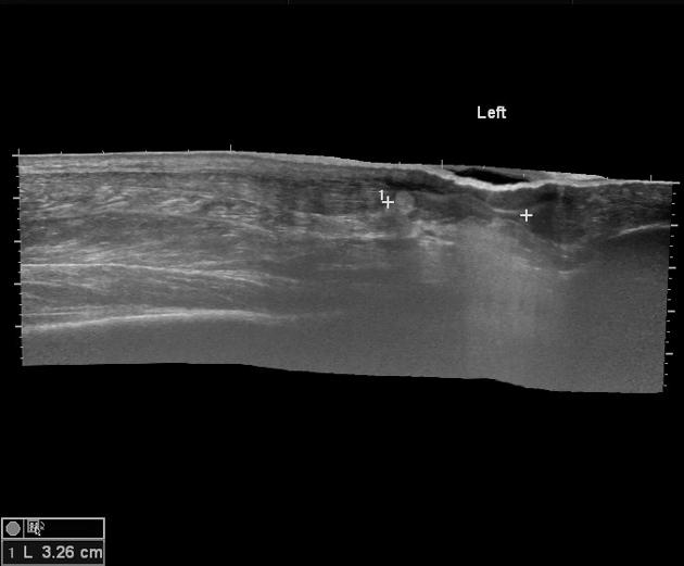
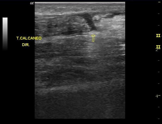
Ultrasound
For partial thickness tears, there is often enlargement of the tendon ( >1 cm) with abnormally hypoechoic or anechoic areas within which correspond to the tear and associated adjacent tendinosis.
For full thickness tears, ultrasound often shows separation of the torn ends with a contour change of the tendon. Also, acoustic shadowing at the margins of the tear from sound beam refraction, and adjacent hypoechoic tendinosis.
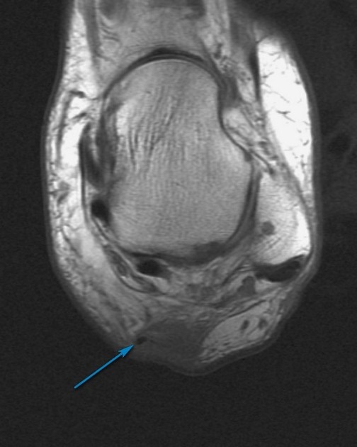
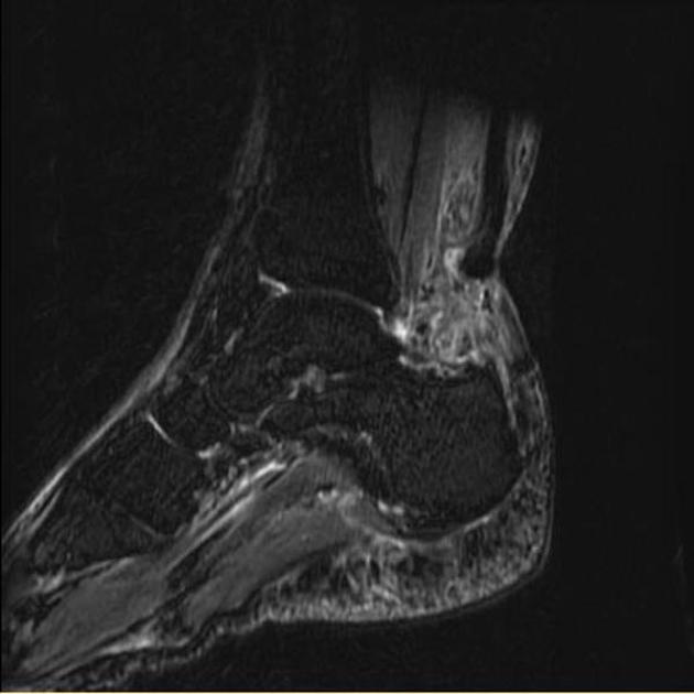
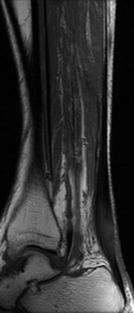
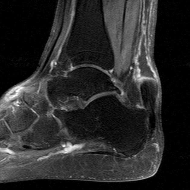
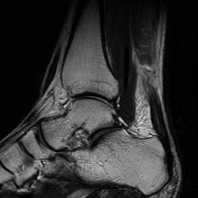
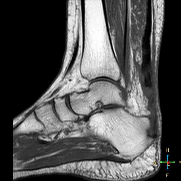
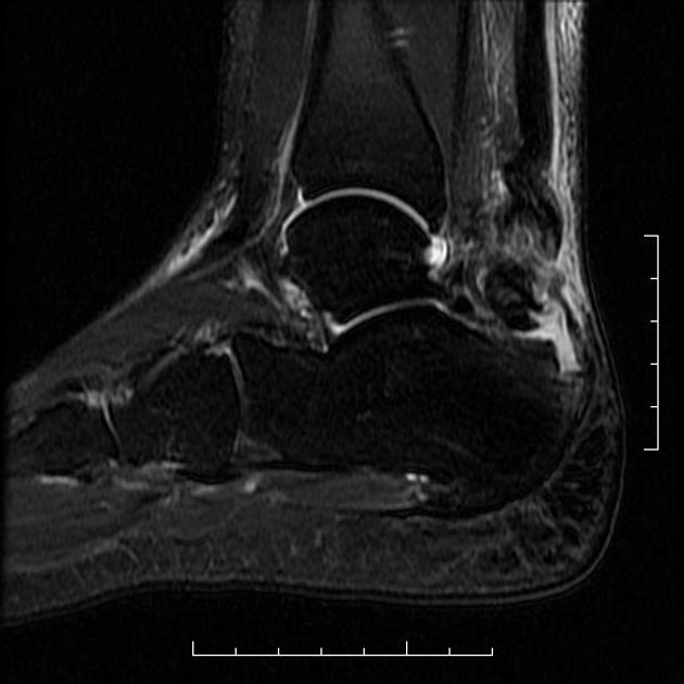
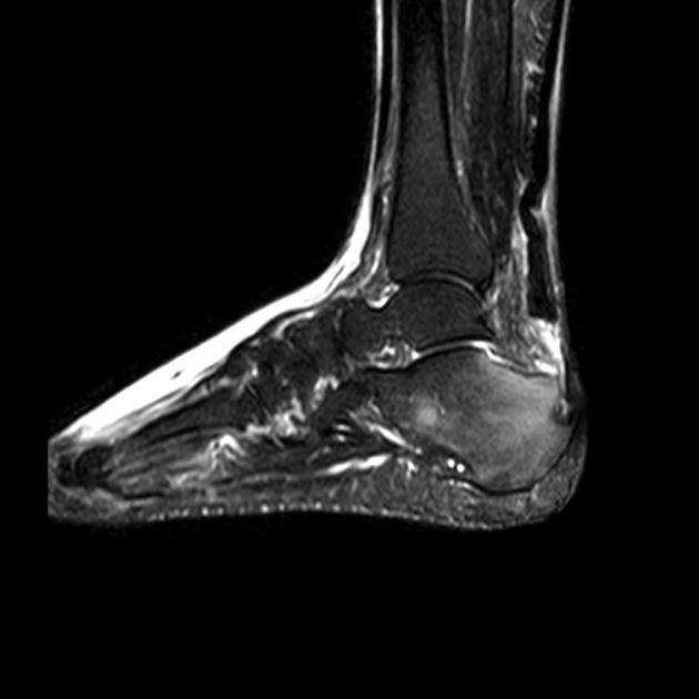
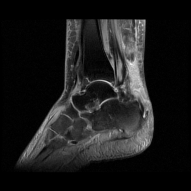
MRI
Appearances can vary:
a full-thickness tear often shows a tendinous gap filled with edema or blood
complete rupture shows retraction of tendon ends
When a plantaris muscle is present then its tendon is usually spared due to its more anterior insertion on the calcaneum.
Signal characteristics
T2: partial thickness or interstitial tears may show high signal on long TR, and tendon swelling to >7 mm AP
Post-operative
post-operative MR imaging may show a tendon gap although this tends to resolve in around 12 weeks 8
post-operatively, Achilles tendon may appear thicker on MR follow up 9
Treatment and prognosis
Treatment depends on the extent of the tear. Partial thickness tears can initially be treated conservatively, with surgery reserved for failure of conservative management, or in some cases for high-performance athletes. Full-thickness tears are normally surgically repaired. If the patient is not deemed suitable for surgical repair (frail, ill, etc.) casting of the ankle in the talipes equinus position may be an alternative.
Surgical repair results in a shorter Achilles tendon and better greater calf muscle strength (less soleus atrophy) than non-surgical treatment 10.
History and etymology
A true rupture of the Achilles tendon was first described by Ambroise Pare in 1575 and first reported in the medical literature in 1633 3.


 Unable to process the form. Check for errors and try again.