
This is a basic article for medical students and other non-radiologists
Acute pancreatitis refers to acute inflammation of the pancreas and is a potentially life-threatening condition.
On this page:
Reference article
This is a summary article; read more in our article on acute pancreatitis.
Summary
- anatomy
-
epidemiology
- epidemiology is dependent on the cause of pancreatitis
- gallstones, idiopathic, alcohol, malignancy, metabolic conditions
- epidemiology is dependent on the cause of pancreatitis
- presentation
-
pathophysiology
- obstruction of the pancreatic ducts is common
- pancreatic enzyme activation results in inflammation
- inflammation of the pancreas causes in interstitial edema and swelling
- continued inflammation results in necrosis, and in some cases hemorrhage
-
investigation
- not required for diagnosis, but may help
- very useful for assessment of complications
-
treatment
- largely supportive, often requiring ICU care in severe cases
- respiratory and cardiovascular support
- careful management of glucose, calcium, and fluid balance
-
prognosis
- dependent on the severity of disease and rapidity of treatment
- complication include necrosis and cyst formation
Role of imaging
- clarify the diagnosis when the clinical picture is confusing
- assess severity and determine prognosis
- detect complications
- determine possible causes
Radiographic features
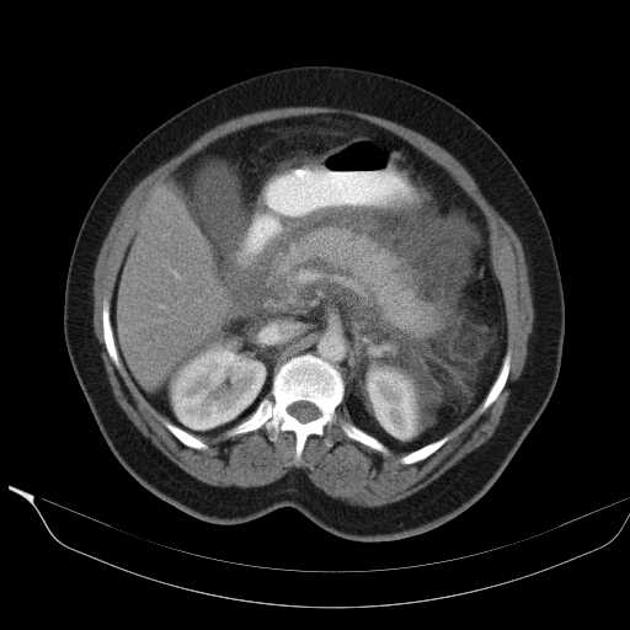
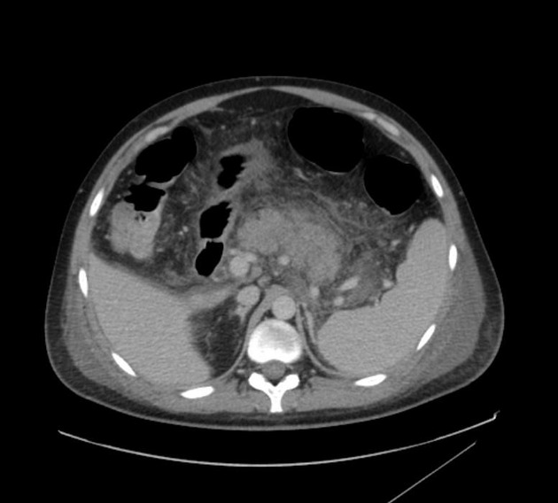
Imaging studies of acute pancreatitis may be normal in mild cases. Contrast-enhanced CT provides the most comprehensive initial assessment, typically with a dual phase (arterial and portal venous) protocol.
CT
Abnormalities that may be seen in the pancreas include:
- typical findings
- focal or diffuse parenchymal enlargement
- changes in density because of edema
- indistinct pancreatic margins owing to inflammation
- surrounding retroperitoneal fat stranding
- necrosis of pancreatic parenchyma
- lack of parenchymal enhancement
- infected necrosis
- difficult to distinguish from aseptic liquefactive necrosis
- the presence of gas is helpful
- abscess formation
- circumscribed fluid collection
- little or no necrotic tissues
- hemorrhage
- high-attenuation fluid in the retroperitoneum or peripancreatic tissues


 Unable to process the form. Check for errors and try again.