
Atrioventricular septal defects (AVSDs), also known as atrioventricular canal defects or endocardial cushion defects, comprise a relatively wide range of defects involving the atrial septum, ventricular septum, and one or both of the tricuspid or mitral valve. They can represent 2-7% of congenital heart defects.
On this page:
Epidemiology
The estimated prevalence is at ~3-4 in 10,000 births.
Associations
Down syndrome (trisomy 21): may be present in up to 50% of cases 4
Edwards syndrome (trisomy 18): may be present in up to 25% of cases 7
-
may be present in up to 10% of cases of asplenia (Ivemark) syndrome 4
Pathology
It results from deficient development of the apical portion of the atrial septum, basal portion of the interventricular septum, as well as the atrioventricular valves. All four chambers of the heart communicate, therefore, both left-to-right and right-to-left shunts can occur.
Classification
Many have been used but can be broadly divided into:
complete
incomplete
An atrioventricular septal defect may also be balanced or unbalanced 8.
Radiographic features
Plain radiograph
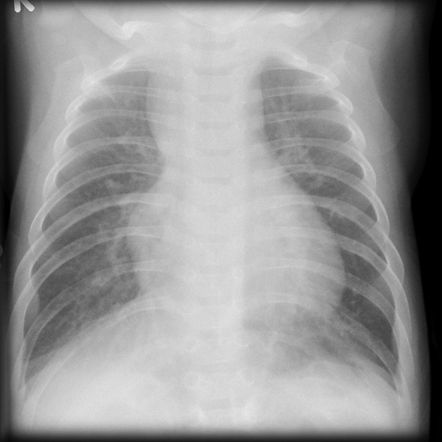
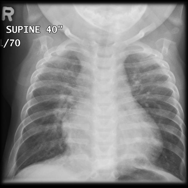
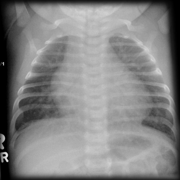
Plain chest radiographic features are often non-specific but may show cardiomegaly +/- features of pulmonary hypertension and mitral valve insufficiency.
Ultrasound
Echocardiography
Allows direct visualization of the defect spectrum; often a large defect of the midline heart structures is seen. Color Doppler often aids in further visualization of the central opening.
Angiography (DSA)
An atrioventricular septal defect can give a classical "Gooseneck" sign on a lateral left ventricular angiogram 3.
MRI
Allows direct visualization of the defect spectrum. Can be superior in assessing cardiac chamber dimensions and the presence/extent of ventricular hypoplasia which is a determinant of surgical risk.
Treatment and prognosis
AVSD closure is generally performed via surgery 9.


 Unable to process the form. Check for errors and try again.