
Atypical lipomatous tumors / well-differentiated liposarcomas (ALT/WDLPS) are locally aggressive adipocytic soft tissue neoplasms and are the most common form of liposarcomas.
On this page:
Terminology
The terms well-differentiated liposarcoma and atypical lipomatous tumor depend on the tumor's localization and resectability. Atypical lipomatous tumors are also termed ‘atypical lipomas’, but this term has been discouraged 1.
Epidemiology
Atypical lipomatous tumors or well-differentiated liposarcomas account for 40-45% of liposarcomas and constitute the largest subgroup of adipocyte malignancies. They occur mostly in adults, with a peak incidence in the 30s and 40s. They are extremely rare in childhood. Inguinal lesions are more common in men; otherwise, there is no gender predominance; however, 1-3.
Associations
Atypical lipomatous tumors/well-differentiated liposarcomas may be associated with Li-Fraumeni syndrome 1.
Diagnosis
The diagnosis of well-differentiated liposarcoma is based on typical pathological features.
Diagnostic criteria
Diagnostic criteria according to the WHO classification of soft tissue and bone tumors (5th edition) differ with subtype 1:
-
lipoma-like subtype:
varying sizes of adipose cells
nuclear atypia in adipose or stromal cells
sclerosing subtype: bizarre hyperchromatic stromal cells in a fibrillary collagenous stroma
inflammatory subtype: atypical stromal cells in a chronic inflammatory infiltrate
The following criteria are described as desirable in challenging cases 1:
MDM2 and/or CDK4 nuclear expression
MDM2 and/or CDK4 gene amplification
Clinical presentation
The most common presentation is that of a painless mass. Deep-seated atypical lipomatous tumors/well-differentiated liposarcomas are most often found incidentally 1.
Complications
If left untreated atypical lipomatous tumors/well-differentiated liposarcomas can dedifferentiate into higher-grade malignancies such as dedifferentiated liposarcoma or undifferentiated pleomorphic sarcoma 1-4.
Pathology
Atypical lipomatous tumors/well-differentiated liposarcomas are adipocytic neoplasms characterized by a proliferation of pleomorphic mature adipocytes of different patterns featuring atypical hyperchromatic stromal cells. They are intersected by fibrous septa and might have myxoid or fibrous components and areas of fat necrosis 1-4.
Location
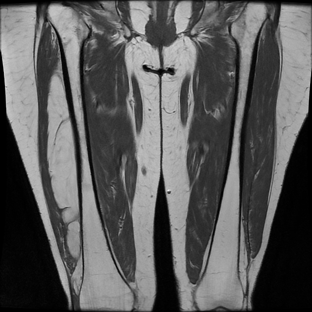
Atypical lipomatous tumors/well-differentiated liposarcomas commonly involve the deep soft tissues in particular of the proximal extremities and the trunk 1-5.
Common locations include the following 1:
Rarer locations of involvement include the following 1:
distal extremities
skin
Macroscopic appearance
Macroscopically atypical lipomatous tumors/well-differentiated liposarcomas usually present as well-circumscribed, large lobulated masses of variable consistency and white to yellowish color. They might display foci of necrosis or small punctate hemorrhages 1.
Microscopic appearance
Atypical lipomatous tumors/well-differentiated liposarcomas can be divided into adipocytic (lipoma-like), sclerosing, and inflammatory morphological subtypes 1-4,6. Larger tumors and especially retroperitoneal tumors commonly show more than one morphologic pattern within the same lesion 1. The microscopic appearance of atypical lipomatous tumors/well-differentiated liposarcomas includes the following features 1,4:
presence of atypical hyperchromatic stromal cells
variable number of lipoblasts
possibly variations in adipocyte size with nuclear atypia (lipoma-like subtype)
possibly copious inflammatory infiltrates (inflammatory subtype)
possibly bizarre stromal cells within a fibrillary sclerotic, collagenous background (sclerosing subtype)
Immunophenotype
Immunohistochemistry stains are usually positive for MDM2 and/or CDK4 1.
Genetics
The pathogenesis of atypical lipomatous tumors/well-differentiated liposarcomas involves MDM2 and/or CDK4 nuclear gene amplification 1,2.
Radiographic features
Imaging features of atypical lipomatous tumors/well-differentiated liposarcomas resemble that of benign lipomas as discussed in the article lipoma vs well-differentiated liposarcoma and include 3-5,7:
size >5 cm or >13 cm 7
thick or nodular septa (>2 mm) with enhancement
multinodular tumor margins
focal nodular patchy non-fatty tissue components
more than 3/4 of fatty tissue
They might displace other organs or tissue.
Ultrasound
Usually appears as multilobulated well-defined mass sometimes with hyperechoic foci 3.
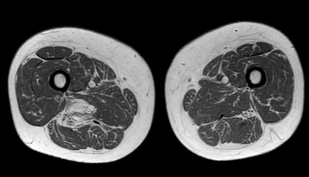
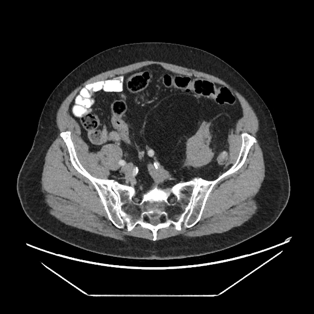
CT
CT usually shows a fat tissue density mass with thick or nodular enhancing septae. Calcifications might be rarely found 4.
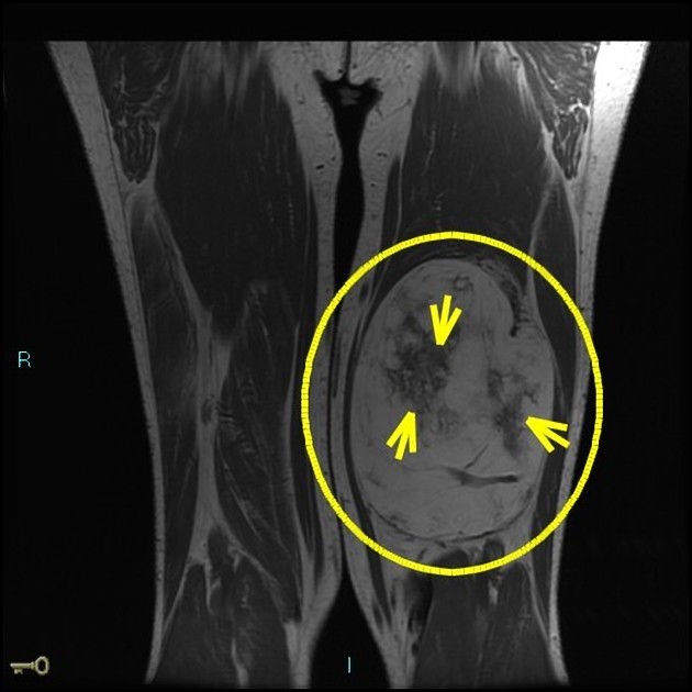
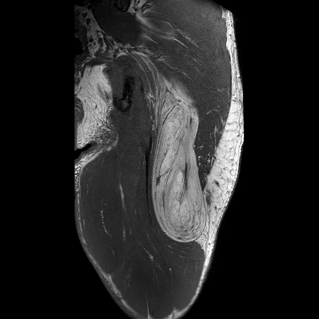
MRI
On MRI atypical lipomatous tumors/well-differentiated liposarcomas will display a mass of fat isointense signal in all sequences. In addition, atypical lipomatous tumors/well-differentiated liposarcomas will show thick septa or nodular non-lipomatous areas with contrast enhancement 3-5.
T1: hyperintense
T2: hyperintense possibly with prominent high signal foci
T2FS/PDFS: hypointense
T1 C+ (Gd): moderate to a marked enhancement of septa
Radiology report
The radiological report should include a description of the following 5,7:
form, location and size
tumor margins
thick septa and septal enhancement
amount of non-adipose tissue
distance from the muscular fascia
relationship to local nerves and vessels
Treatment and prognosis
The management of choice in atypical lipomatous tumors/well-differentiated liposarcomas is resection, which is curative if complete 1. These tumors are not sensitive to radiotherapy or chemotherapy 2. Tumor prognosis and the likelihood of distant metastases depend on the risk of dedifferentiation and on how amenable the location is to surgical excision. It varies with the location of the tumor ranging from >20% in the retroperitoneum to <2% in the extremities. The tumors do not cause distant metastasis unless there is dedifferentiation.
Tumors of the mediastinum retroperitoneum or spermatic cord have a worse prognosis, due to their location they are also more prone to local recurrence 1,2. On the other hand patients with tumors affecting the extremities have a very good prognosis with >90% survival after 10 years of follow-up and thus the name atypical lipomatous tumor 8. For selected patients with atypical lipomatous tumors of the extremities, active surveillance has been suggested as a viable option to prevent overtreatment 8.
History and etymology
Liposarcomas were first reported by the German pathologist Rudolph Virchow in 1857 who called them "myxoma lipomatodes malignum" 9,10. After the 1950s a further classification of liposarcoma into the well-differentiated, myxoid and dedifferentiated was suggested 11-15. The terms atypical lipoma or atypical lipomatous tumor of which the former is no longer recommended 1 have been introduced by the American pathologist Harry L Evans in 1979 14,15.
Differential diagnosis
Conditions or tumors which can mimic the presentation and/or the appearance of atypical lipomatous tumors/well-differentiated liposarcomas include 1-5:
fat necrosis


 Unable to process the form. Check for errors and try again.