
Carotid artery stenosis, also known as internal carotid artery stenosis or extracranial carotid artery stenosis, is usually caused by an atherosclerotic process and is one of the major causes of stroke and transient ischemic attack (TIA).
This article refers to stenosis involving the carotid bulb and the proximal segment of internal carotid artery (ICA), as both are the most common sites of symptomatic and clinically relevant stenosis.
On this page:
Epidemiology
Carotid artery stenosis is usually seen in the elderly and more commonly in males. In asymptomatic patients over the age of 80, approximately 3% of men and 1% of women will have severe stenosis 9.
Atherosclerotic carotid arterial disease accounts for ~15% of all ischemic strokes and TIAs 6,7. The annual incidence of extracranial carotid artery stenosis as a cause of stroke accounts for ~13 per 100,000 population in the USA 8.
Clinical presentation
Carotid artery stenosis can result in wide-ranging symptoms from ischemic stroke or transient ischemic attack 7. Rarely, severe carotid artery stenosis can result in convexal subarachnoid hemorrhage due to rupture of compensatory pial vessels 10.
Pathology
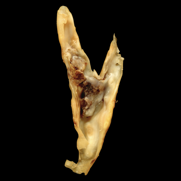
The plaques formed in the carotid vessels can be divided into four types:
type I: predominantly hemorrhage, lipid, cholesterol, and proteinaceous material
type II: dense fibrous connective tissue with >50% volume of hemorrhage, lipid, cholesterol, and proteinaceous material
type III: dense fibrous connective tissue with <50% volume of hemorrhage, lipid, cholesterol, and proteinaceous material
type IV: dense fibrous connective tissue
Radiographic features
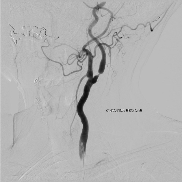
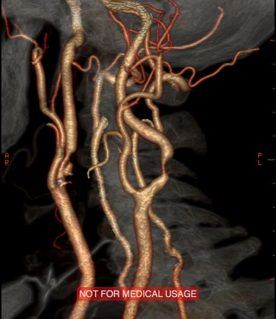
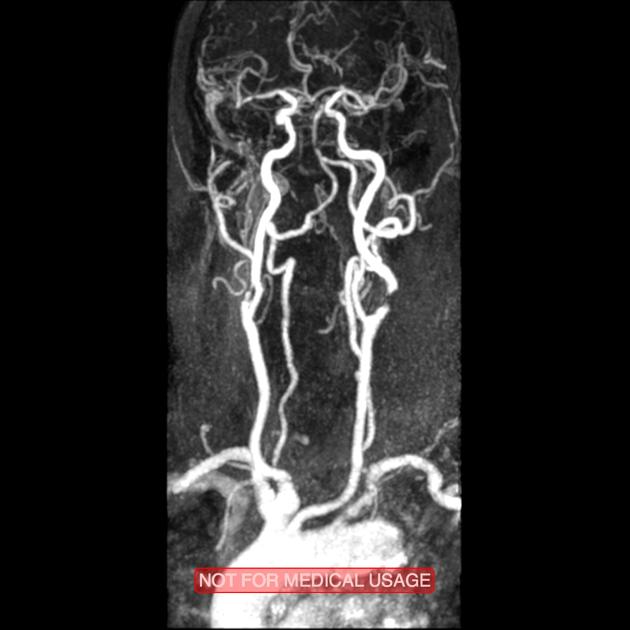
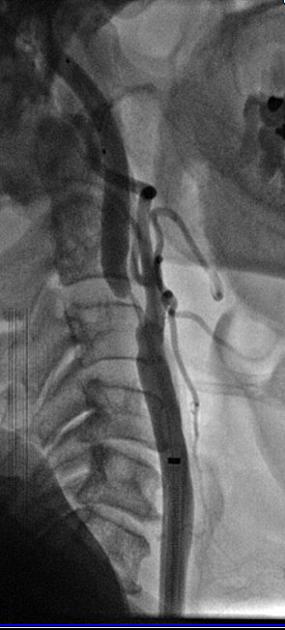
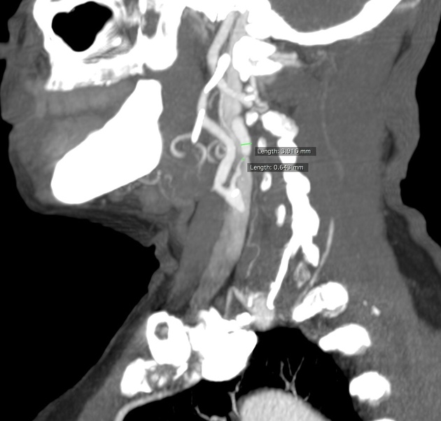
Conventional angiography has been considered the standard method for evaluating carotid stenosis, moreover, the trials published in the 1990s (NASCET 11, ECST 12, and ACAS 13) were based on this method. However, the introduction and development of ultrasound Doppler, CT angiography (CTA), and MRI angiography (MRA) have been replacing angiography for diagnostic purposes, essentially reserving it for endovascular treatment.
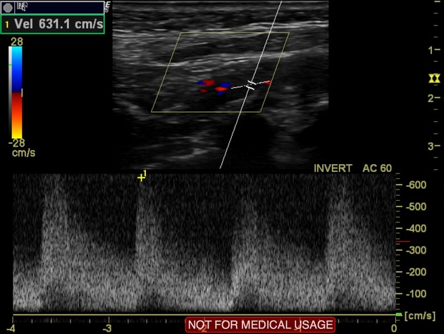
Ultrasound
On grayscale, characterization of plaques can be performed:
type I: predominantly hypoechoic with thin echogenic rim
type II: echogenic plaque with >50% hypoechoic areas
type III: echogenic plaque with <50% hypoechoic areas
type IV: uniformly echogenic plaque
They can be termed as:
homogeneous (type I and IV)
heterogeneous (type II and III)
Doppler ultrasound has become the first choice for carotid stenosis screening, permitting the evaluation of both the macroscopic appearance of plaques as well as flow characteristics 5. Hemodynamically significant carotid stenosis is usually referred to a further CTA or MRA study.
Angiography (DSA)
The North American Symptomatic Carotid Endarterectomy Trial (NASCET) demonstrated a conclusive benefit for carotid endarterectomy in patients with symptomatic 70-99% ICA stenosis 2. NASCET was established by angiographic calculation of ICA stenosis percentage using the following formula:
% ICA stenosis = (1 - [narrowest ICA diameter/diameter normal distal cervical ICA]) x 100
The European Carotid Surgery Trial (ECST) also demonstrated benefits for carotid endarterectomy in patients with symptomatic higher than 80% ICA stenosis 3. ECST was established by angiographic calculation of ICA stenosis percentage using the following formula:
% ICA stenosis = (1 - [diameter of the most stenotic part/estimated original diameter at the site of the stenosis]) x 100
Treatment and prognosis
NASCET and ECST trials proved the benefits of performing endarterectomy in those patients with symptomatic high-grade stenosis. In 1995 the Asymptomatic Carotid Atherosclerosis Study (ACAS) demonstrated that patients with asymptomatic carotid artery stenosis of 60% or greater reduction in diameter benefited from endarterectomy, having a reduced 5-year risk of ipsilateral stroke 4.
Procedures used to treat carotid stenosis include carotid endarterectomy and carotid arterial stenting.
Differential diagnosis
External links
If any of these links are broken or for other problems and questions, please contacteditors@radiopaedia.org.


 Unable to process the form. Check for errors and try again.