
Cerebral venous thrombosis (CVT) (plural: thromboses) refers to the occlusion of venous channels in the cranial cavity, including dural venous thrombosis, cortical vein thrombosis and deep cerebral vein thrombosis. They often co-exist and the clinical presentation among them is very similar and non-specific. Furthermore, diagnostic imaging features can be subtle.
On this page:
Epidemiology
Cerebral venous thrombosis is a rare condition accounting for approximately 0.5% of all cases of cerebrovascular disease worldwide 8. Demographics of affected patients reflects underlying predisposing factors, which are identified in the majority of cases (87.5%) with many patients having more than one coexistent risk factors 2:
-
hormonal
exogenous
pregnancy
puerperium
-
steroids
oral contraceptive pill: very common cause in female patients <50 years of age 2
COVID-19 vaccine, especially AstraZeneca 10
-
prothrombotic hematological conditions: 35% 2
-
local factors
skull abnormalities/trauma
compressing mass: e.g. meningioma
infection: especially mastoid sinus (dural sinus occlusive disease - DSOD)
-
systemic illness
dehydration: e.g. gastroenteritis
malignancy
idiopathic: ~12% 2
Clinical presentation
Unlike most other intracranial vascular conditions, the presentation can be highly variable and range from asymptomatic, seizure to coma and death, and may mimic a host of other conditions 1.
Symptoms
headaches (89-91%) 8
decreased/altered conscious state
decreased/altered vision
nausea and vomiting
Signs
cranial nerve palsies
focal neurological deficits (52-68%) 8
seizures (39-44%) 8
coma
Pathology
Cerebral venous thrombosis pathogenesis remains poorly understood 5. There is an extensive list of known risk factors, already mentioned above.
The lesion volume is related to the development of collateral veins in the affected venous segment. Venous hypertension from a poor outflow can lead to edema, cerebral venous infarction (~50% of cases 1) and even hemorrhage.
Superior sagittal sinus or the dominant transverse sinus thrombosis can affect the arachnoid granulations absorption of cerebrospinal fluid, a consequent increase of cerebral swelling may occur 1.
Radiographic features
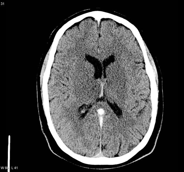
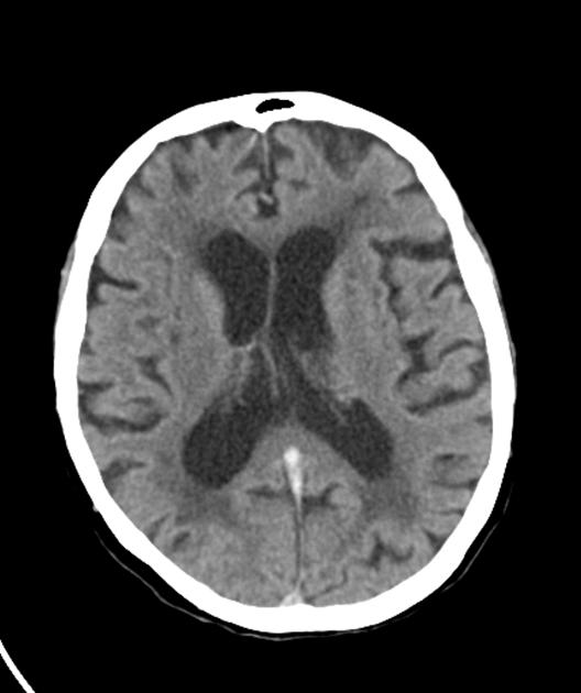
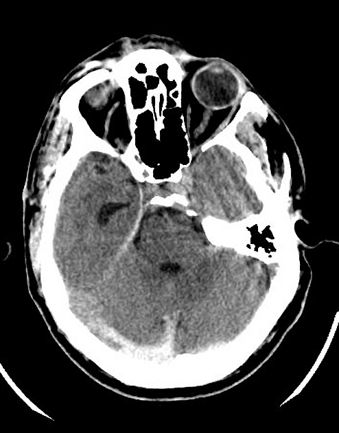
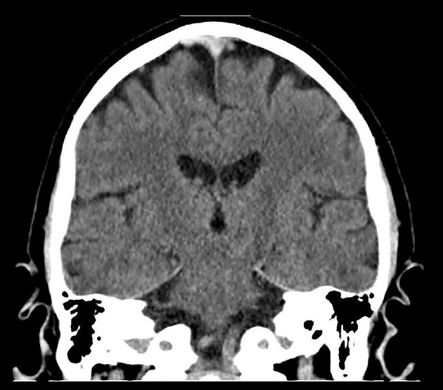
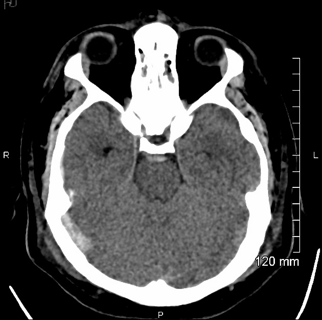
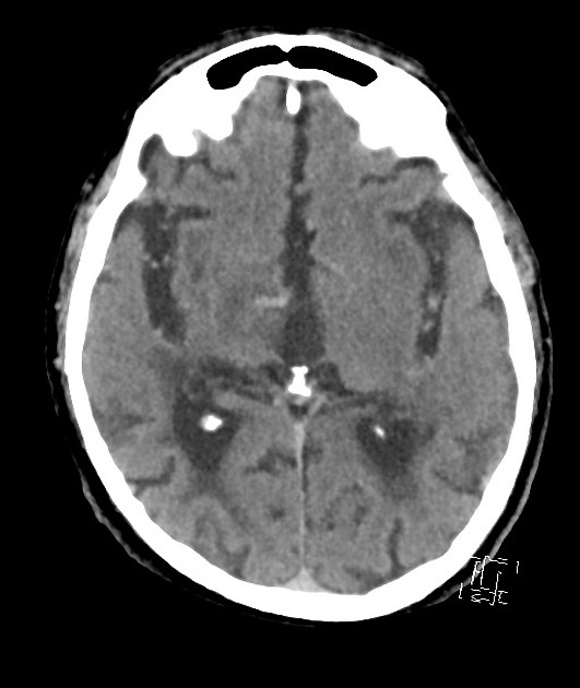
Unenhanced CT is usually the first imaging investigation performed given the nonspecific clinical presentation in these cases.
CT
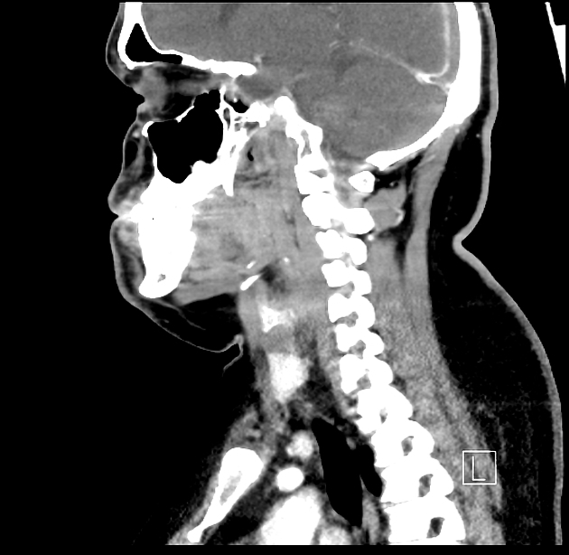
Non-contrast CT, when not associated with venous hemorrhage or infarction can be a subtle finding, relying on hyperdensity of the sinus being identified 1,5. Thrombus can appear as a hyperdense vein or sinus for the first 7-14 days; this is an accurate sign when present 6. The cashew nut sign, which describes small, concave, juxtacortical intracerebral hemorrhages, is specific for cerebral venous thrombosis when present 14.
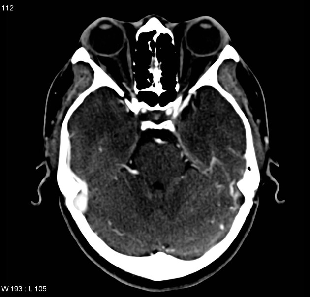
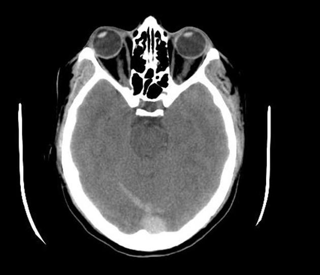
With contrast administration, especially with a CT venogram, a filling defect in a sinus is sought. When in the superior sagittal sinus it is referred to as the 'empty delta sign'. CTV has a reported sensitivity of 95% compared to DSA as the gold standard 1.
Filling defects should not be confused with Pacchionian bodies (arachnoid granulations) which can be seen in essentially all dural sinuses and are especially common in the superior sagittal sinus and transverse sinus.
CT perfusion
Although not used routinely in clinical practice, whole brain CT perfusion may assist in establishing the diagnosis of CVT by detecting perfusion abnormalities that do not correspond with arterial territories 9.
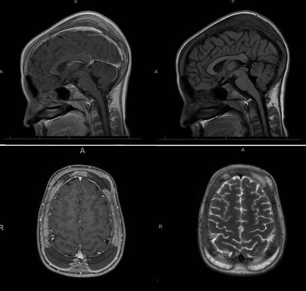
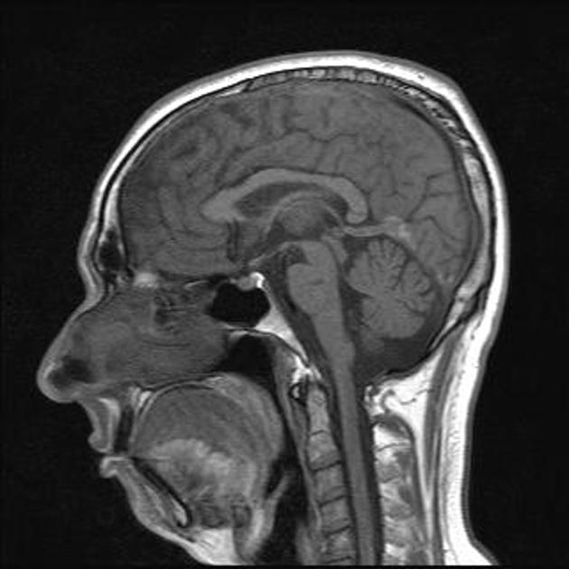
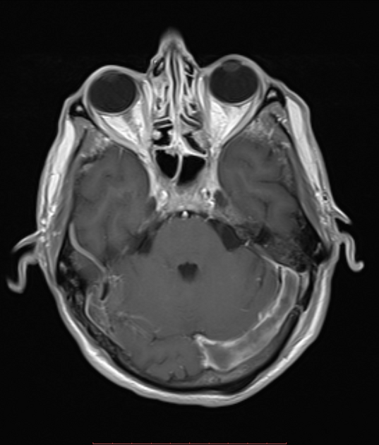
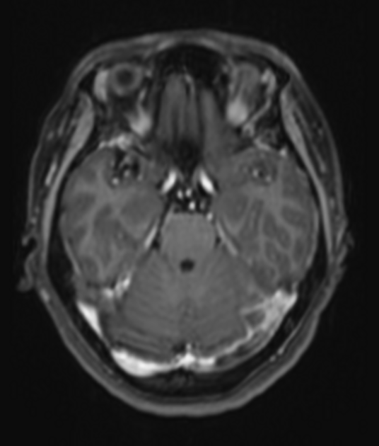
MRI
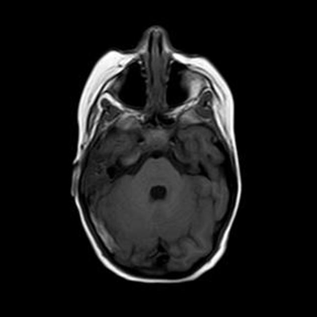
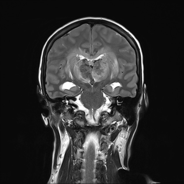
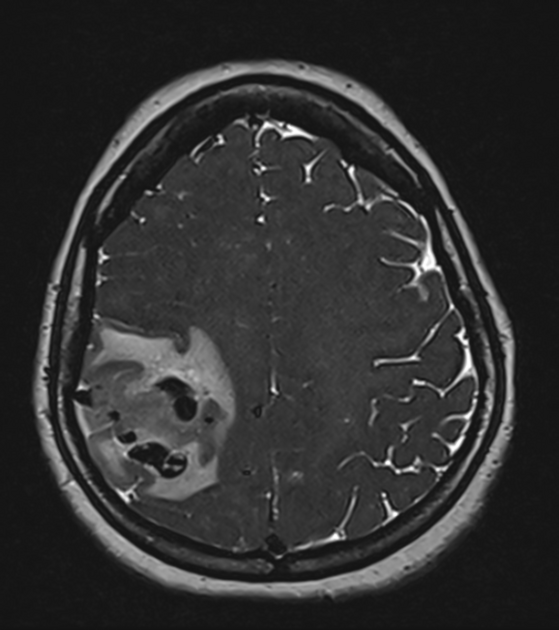
MRI is able to both visualize the clot as well as the sequelae.
The clot acutely is isointense on T1 and hypointense on T2 (this can mimic a flow void), with subacute clot becoming hyperintense on T1.
Cerebral edema can be identified even in the absence of neurological dysfunction or infarction 1.
-
FLAIR
a sulcal hyperintensity may reflect the presence of a localized subarachnoid hemorrhage
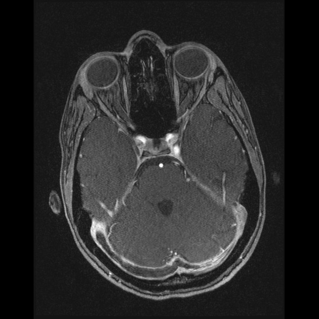
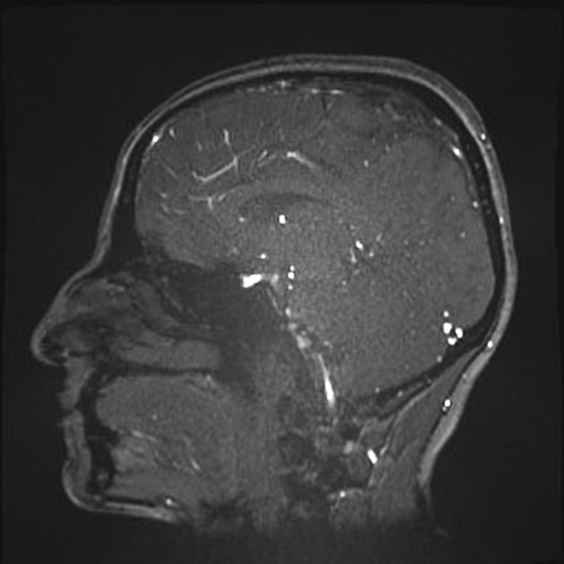
-
T1 C+
may demonstrate focal pachymeningeal and leptomeningeal enhancement due to elevated blood pressure upstream of the thrombosis
-
DWI/ADC
the clot may demonstrate diffusion restriction on chronic stages
-
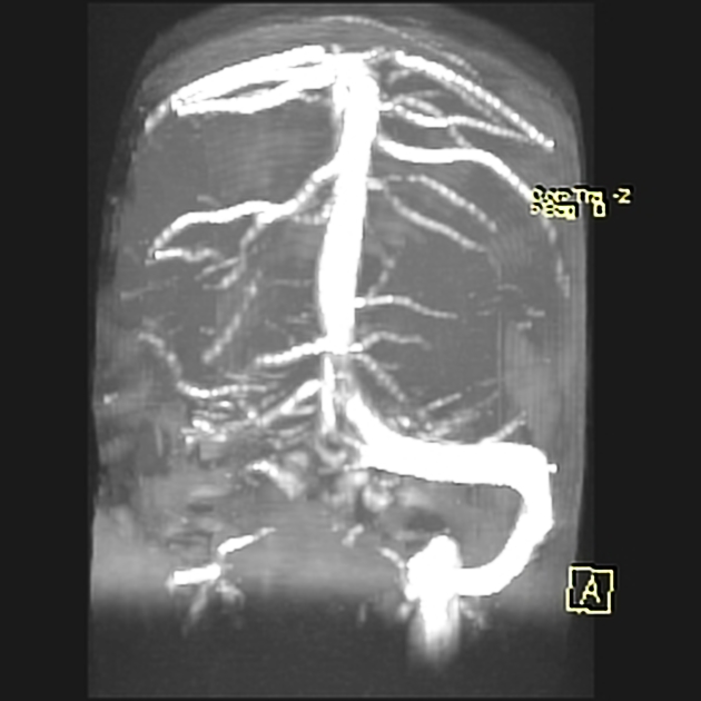
will demonstrate a lack of flow. 2D time of flight (TOF) venography is routinely performed in suspected cases. Contrast MR venography is more sensitive in detecting dural venous sinus thrombosis than TOF venography. Hypoplastic dural sinuses and low flow areas remain a major problem with 2D TOF.
Angiography (DSA)
Although digital subtraction angiography (DSA) has historically been the gold standard, the relative lack of experienced angiographic skills and invasive nature of the examination has led to a dramatic decline in its use as a primary mode of diagnosing cerebral venous thrombosis.
Treatment and prognosis
In ~50% of cases, cerebral venous thrombosis progresses to venous infarction 1. Unlike arterial infarcts, venous infarcts usually present after some days 1:
<2 days of symptom onset: 30%
2-30 days: 50%
>30 days: 20%
The mainstay of hyperacute treatment is heparin, even in the setting of hemorrhagic venous infarction 2,4. Once the patient is stable, alternative enteral anticoagulation is commenced, including direct oral anticoagulants or warfarin 11. The optimal duration of anticoagulation is unknown, but generally at least 3 months 12.
Surgical interventions are considered on a case-by-case basis. Interventional neuroradiologists/neurosurgeons can perform catheter-directed thrombolysis by using targeted thrombolytics in the affected sinuses. In patients who are critically unwell with pending brain herniation, observational data, in the absence of randomized control trials, suggests improved outcomes in selected patients managed with decompressive craniectomy 13.
The natural history of cerebral venous thrombosis is highly variable, with some patients having minimal or no symptoms and an uneventful recovery (~65%), whereas others have a fulminant course culminating in extensive venous infarction and dependency or death (~20%) 2.
Not surprisingly, hemorrhagic venous infarcts and co-existing malignancy correlate with poor outcome 2. Deep cerebral venous thrombosis also has a negative impact on prognosis due to the usually bilateral involvement of the thalami 3.
Complications
Dural arteriovenous fistula, increased CSF pressure, and subarachnoid hemorrhage have been reported as possible complications after cerebral venous thrombosis.
Differential diagnosis
The main differentials of cerebral vein thrombosis are:
hypoplastic venous sinuses


 Unable to process the form. Check for errors and try again.