
Clear cell meningiomas are a histological variant of meningioma with poorer prognosis and a higher rate of recurrence. In the 5th Edition of the WHO classification of CNS tumors, they are classified as grade 2 tumors, regardless of mitotic index, cellular atypia/anaplasia, or presence of brain invasion.
On this page:
Epidemiology
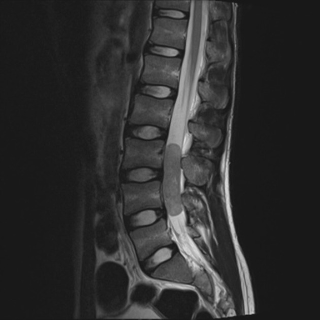
Clear cell meningiomas have been reported to occur at a younger age group (mean age of ~30 years) and are more frequently located within the spinal canal (see spinal meningioma) and posterior fossa 1-3. No convincing predilection for either gender has been reported 1.
Clinical presentation
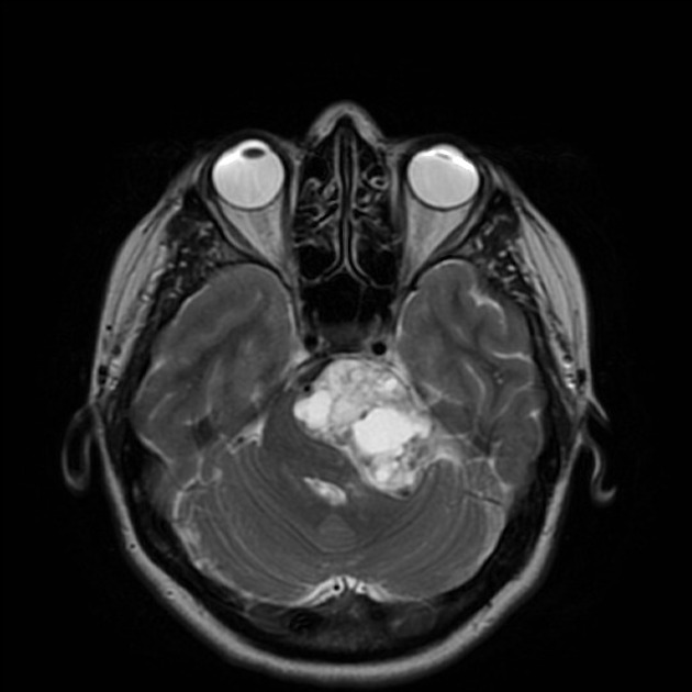
Although generally, they present in a similar fashion to other meningiomas, their predilection for the posterior fossa and spinal canal clearly has implications for the presenting symptoms.
Pathology
Clear cell meningiomas appear similar to meningothelial (syncytial) meningiomas. However, the tumor cells have vacuolated cytoplasm; thus the moniker 'clear cell' 1. Histologically these tumors resemble other clear cell tumors such as oligodendroglioma, hemangioblastoma, germinoma, pleomorphic xanthoastrocytoma (PXA), clear-cell ependymoma and metastases from renal cell carcinoma 1.
Radiographic features
Radiographic features are similar to those of the more common 'typical' meningiomas and are thus not repeated here.
Treatment and prognosis
Despite benign-appearing histology, clear cell meningiomas have a tendency to recur locally (higher than 60% in some series, and particularly high in intracranial disease) and metastasize within the CSF space 1-3.


 Unable to process the form. Check for errors and try again.