
Cockayne syndrome is a rare autosomal recessive dysmyelinating disease. Cockayne syndrome is classified among the childhood leukodystrophies, and brain imaging findings are cardinal features suggesting the diagnosis of Cockayne syndrome. Previously published Cockayne syndrome imaging studies have described major brain atrophy (predominantly white matter), bilateral and symmetrical calcifications, and a lack of myelination of the white matter, but whether these findings are due to hypomyelination or demyelination remains unclear.
On this page:
Clinical presentation
Clinical features include failure to thrive, neurodevelopmental delay, cutaneous photosensitivity, pigmentary retinopathy, sensorineural hearing loss, dental caries, and cachectic dwarfism.
The diagnosis is considered very likely if the first 2 clinical criteria and at least 3 of the other criteria mentioned above are present.
Pathology
Genetics
Cockayne syndrome is an autosomal recessive disorder due to mutations in genes encoding nucleotide excision repair proteins ERCC6 (CSA) and ERCC8 (CSB).
Cockayne syndrome belongs to the group of nucleotide excision repair disorders that includes xeroderma pigmentosum. In some variants, xeroderma pigamentosum can occur in combination with Cockayne syndrome.
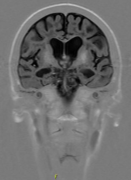
Radiographic features
CT
Cockayne syndrome is one of the causes of basal ganglia calcifications in a child. Calcification may also occur in cerebellar and cerebral cortical regions. CT may also show early atrophy.
MRI
There is atrophy which predominantly involves the supratentorial white matter, the cerebellum, the corpus callosum, and the brainstem 1.
-
T2:
calcification may be seen as low signal in putaminal (most common), dentate nuclear and cortical regions
tigroid pattern in the deep white matter, with hypointense foci within hyperintensity 1
-
MRS:
The combination of demyelination and basal ganglia calcification may, therefore, be helpful in the imaging of this entity 3.
History and etymology
This condition is named after Edward Alfred Cockayne, English physician (1880-1956).
Differential diagnosis
Pelizaeus-Merzbacher disease: may show similar features in early stages, but no calcification occurs
Mitochondrial diseases: the severe and progressive brain atrophy is more suggestive of Cockayne syndrome
congenital cytomegalovirus infections: brain calcifications typically show a periventricular subependymal distribution
Aicardi-Goutières syndrome: calcifications punctuate in basal ganglia, deep and periventricular white matter


 Unable to process the form. Check for errors and try again.