
Conjoined twin pregnancy is a rare occurrence resulting from the failure of a zygote to separate completely after 13 days 11. This results in the twins being physically joined.
On this page:
Epidemiology
The prevalence of conjoined twins ranges from 1:50,000 to 1:200,000. They are more common in parts of Southeast Asia and Africa with prevalence rates as high as 1:14,000 to 1:25,000. There is a recognized female predisposition (F: M of approximately 3:1).
Pathology
Embryology
Conjoined twins are monozygotic, monoamniotic, and monochorionic (MCMA) (see multifetal pregnancy) and result due to a failure of normal complete separation of the embryonic plate from an incomplete delayed division of the inner cell mass. This is thought to occur around 13-17 days of gestation.
Classification
Conjoined twins are classified according to the most prominent site of interconnection
Ventral union
cephalopagus: face (rare)
thoracopagus: thorax, commonest site (~70% 2,9)
omphalopagus, xiphopagus: abdomen
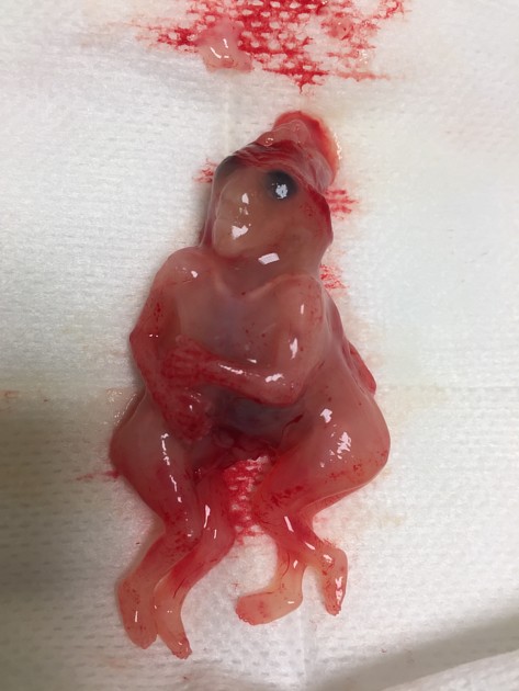
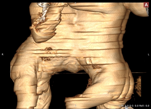
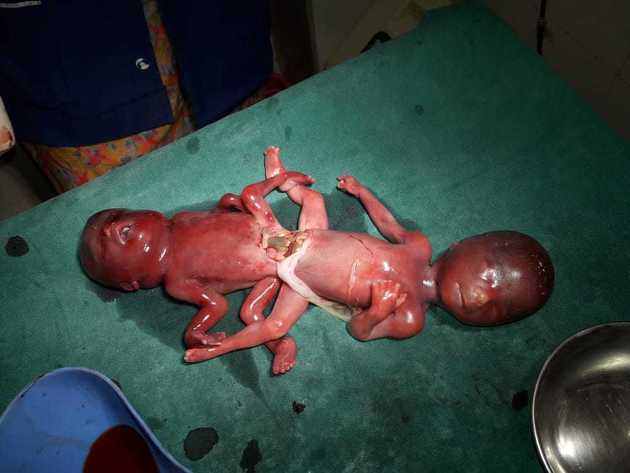
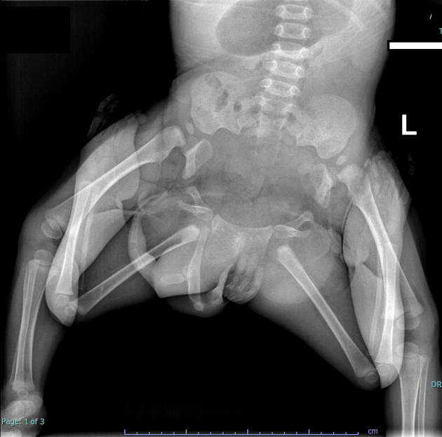
ischiopagus: pelvis ~5%
Dorsal union
craniopagus: skull (rare)
pygopagus: sacrum
rachipagus: vertebral fusion above the sacrum
Lateral union
If more than one area is connected the terms are combined, e.g. thoraco-omphalopagus (thoracic and abdominal fusion).
Other descriptive terms include:
diprosopus: two faces with one head and body
dicephalus: two heads with one body
syncephalus: facial fusion with or without thoracic fusion
Associations
There is a higher incidence of congenital malformations in conjoined twins (10-20%) which are unrelated to the point of fusion such include
Radiographic assessment
Ultrasound
Thus identification of a dividing membrane or two placentas excludes the diagnosis. Definitive sonographic features will depend on the type of fusion.
General features include:
lack of a separating inter-twin membrane
non-separable skin contours with an inability to separate the fetal bodies
detection of other anomalies in a twin gestation
solitary umbilical cord with more than 3 vessels present
both fetal heads persistently at the same level
backward flexion of the cervical spine (due to the fact that most conjoined twins are fused ventrally and face each other
bibreech or less commonly, bicephalic presentation
constant relative fetal positions
Treatment and prognosis
The prognosis for conjoined twins, in general, is quite poor. Approximately 40-60% of conjoined twins are stillborn and almost 35% of live births do not survive beyond 24 hours. Of those who do survive, surgical separation is sometimes possible (but with higher failure rates if performed within the first 3 weeks 9). Surgical separation, in general, is in most cases very challenging with high mortality, depending on the complexity of shared structures. Of those with thoracopagus, ~75% have extensively joined hearts which in turn preclude a successful separation.


 Unable to process the form. Check for errors and try again.