
Early pregnancy roughly spans the first ten weeks of the first trimester.
On this page:
Radiographic features
Ultrasound
0-4.3 weeks: no ultrasound findings
-
4.3-5.0 weeks:
possible small gestational sac
possible double decidual sac sign (DDSS)
possible intradecidual sac sign (IDSS)
-
5.1-5.5 weeks:
gestational sac should be visible by this time
-
5.5-6.0 weeks
yolk sac should be visible by this time
gestational sac should be ~6 mm in diameter
-
>6.0 weeks
fetal pole may be identifiable on endovaginal ultrasound (1-2 mm)
fetal heart rate (FHR) should be ~100-115 bpm
gestational sac should be ~10 mm in diameter
-
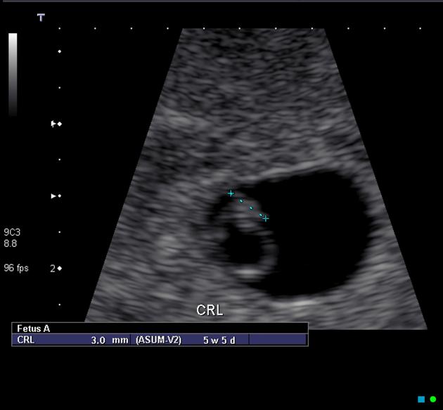
6.5 weeks
crown rump length (CRL) should be ~5 mm
-
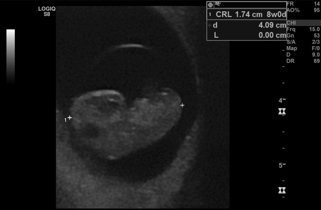
7-8 weeks
CRL is between 11-16 mm
cephalad and caudal poles can be identified
-
8-9 weeks
CRL is between 17-23 mm
limb buds appear
head can be seen as separate from the body
-
9-10 weeks
CRL is between 23-32 mm
fetal heart rate 170-180 bpm
fetal movement can be seen
a round hypoechoic structure in the fetal brain represents a developing embryonic/fetal rhombencephalon
nuchal translucency may begin to be seen
Transvaginal/endovaginal (TV/EV) scanning
intradecidual sac sign (IDSS): early sign on a TV scan
when the MSD measures 25 mm, an embryo must be visible
when the CRL measures >7 mm, an embryo must show cardiac activity
an embryo should be seen <=14 days after a scan with a gestational sac without a yolk sac
an embryo should be seen <=11 days after a scan with a gestational sac and a yolk sac
Transabdominal (TA) scanning
when the MSD measures 20 mm a yolk sac should be visible
when the MSD measures 25 mm, an embryo must be visible
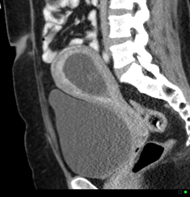
CT/MRI
Occasionally, early pregnancy is unintentionally imaged by CT or sometimes MRI is done for some concurrent pathology, and its important to know the imaging findings 3.
fluid-filled cystic structure in endometrial cavity (well identified on MRI, and may be visible on CT especially on delayed post-contrast images)
developing placenta seen as curvilinear enhancing structure
fetal pole may be seen in delayed first trimester imaging
-
corpus luteal cyst may be visible in one of the ovaries
unilocular <3 cm cyst with irregular crenated and enhancing walls
Differential diagnosis to be considered with a positive urinary pregnancy test includes
If urinary pregnancy test is negative similar findings may suggest submucosal fibroid or retained products of conception.
Practical points
The earlier in pregnancy a scan is performed, the more accurate the age assignment from crown rump length. The initial age assignment should not be revised on subsequent scans 5.
Overall, the accuracy of sonographic dating in the first trimester is ~5 days (95% confidence range).


 Unable to process the form. Check for errors and try again.