
Epiploic appendage
Citation, DOI, disclosures and article data
At the time the article was created Henry Knipe had no recorded disclosures.
View Henry Knipe's current disclosuresAt the time the article was last revised Hossein Salehzadeh had no financial relationships to ineligible companies to disclose.
View Hossein Salehzadeh's current disclosures- Appendices epiploicae
- Epiploic appendages
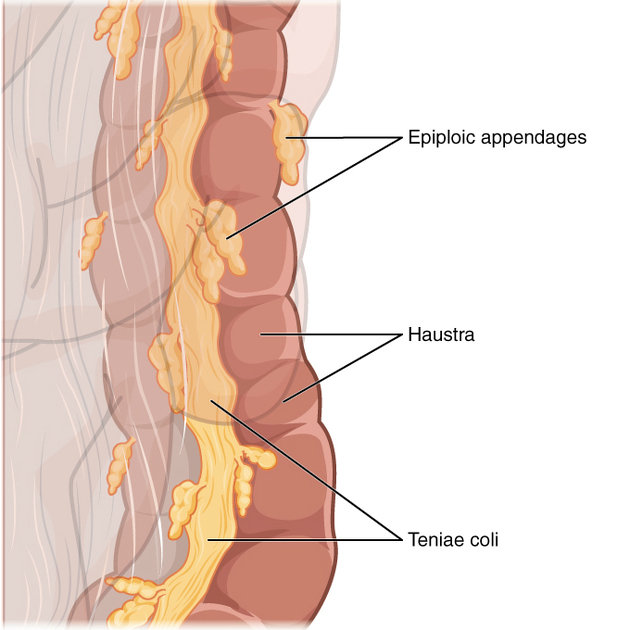
Epiploic appendages (or appendix epiploica, plural: appendices epiploicae) are peritoneum-lined protrusions of subserosal fat that arise from the surface of the large bowel.
On this page:
Gross anatomy
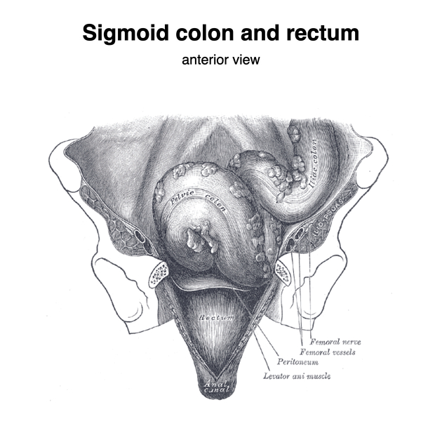
Epiploic appendages typically measure 1.5 x 3.5 cm but have been reported to measure up to 15 cm in length 4. There are between 50-100 of them in the large bowel, from the caecum (where they may be absent) to the rectosigmoid junction. They are distributed longitudinally in two rows on the medial (along the taenia libera) and the posterolateral (along the taenia omentalis) aspects of the large bowel. There is only one row of epiploic appendages along the transverse colon and there are none along the rectum.
Blood supply
They are supplied by one to two small nutrient arteries that pierce the bowel serosa; it is here where colonic diverticula are thought to arise.
Radiographic features
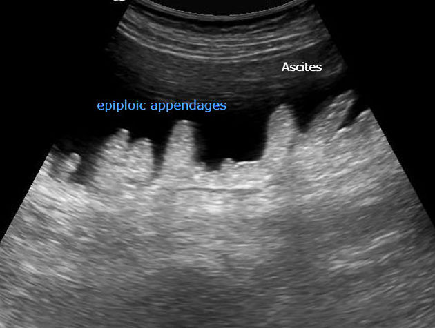
Not normally visible on fluoroscopy, radiography or CT, unless they are surrounded by contrasting material (e.g. haemoperitoneum, ascites, or contrast medium from hysterosalpingography) 4.
Related pathology
Quiz questions

References
- 1. Schuenke M, Schulte E, Schumacher U et-al. Neck and Internal Organs. Thieme. ISBN:1604062940. Read it at Google Books - Find it at Amazon
- 2. Rashid A, Nazir S, Hakim SY et-al. Epiploic appendagitis of caecum: a diagnostic dilemma. Ger Med Sci. 2013;10: Doc14. doi:10.3205/000165 - Free text at pubmed - Pubmed citation
- 3. Abdominal wall hernias. Springer. ISBN:0387950044. Read it at Google Books - Find it at Amazon
- 4. Ghahremani GG, White EM, Hoff FL et-al. Appendices epiploicae of the colon: radiologic and pathologic features. Radiographics. 1992;12 (1): 59-77. doi:10.1148/radiographics.12.1.1734482 - Pubmed citation
Incoming Links
- Epiploic appendagitis
- Epiploic appendagitis
- Rectum and anus (Gray's illustrations)
- Epiploic appendagitis
- Epiploic appendagitis
- Epiploic appendagitis
- Epiploic appendagitis
- Epiploic appendagitis
- Epiploic appendagitis
- Epiploic appendagitis
- Epiploic appendagitis
- Epiploic appendagitis
- Epiploic appendagitis
- Epiploic appendagitis
- Epiploic appendagitis
- Epiploic appendagitis
- Epiploic appendagitis mimicking acute appendicitis
- Epiploic appendages (US)
- Acute epiploic appendagitis
- Epiploic appendagitis
Related articles: Anatomy: Abdominopelvic
- skeleton of the abdomen and pelvis
- muscles of the abdomen and pelvis
- spaces of the abdomen and pelvis
- anterior abdominal wall
- posterior abdominal wall
- abdominal cavity
- pelvic cavity
- perineum
- abdominal and pelvic viscera
- gastrointestinal tract
- spleen
- hepatobiliary system
-
endocrine system
-
adrenal gland
- adrenal vessels
- chromaffin cells
- variants
- pancreas
- organs of Zuckerkandl
-
adrenal gland
-
urinary system
-
kidney
- renal pelvis
- renal sinus
- avascular plane of Brodel
-
variants
- number
- fusion
- location
- shape
- ureter
- urinary bladder
- urethra
- embryology
-
kidney
- male reproductive system
-
female reproductive system
- vulva
- vagina
- uterus
- adnexa
- Fallopian tubes
- ovaries
- broad ligament (mnemonic)
- variant anatomy
- embryology
- blood supply of the abdomen and pelvis
- arteries
-
abdominal aorta
- inferior phrenic artery
- coeliac artery
- superior mesenteric artery
- middle suprarenal artery
- renal artery (variant anatomy)
- gonadal artery (ovarian artery | testicular artery)
- inferior mesenteric artery
- lumbar arteries
- median sacral artery
-
common iliac artery
- external iliac artery
-
internal iliac artery (mnemonic)
- anterior division
- umbilical artery
- superior vesical artery
- obturator artery
- vaginal artery
- inferior vesical artery
- uterine artery
- middle rectal artery
-
internal pudendal artery
- inferior rectal artery
-
perineal artery
- posterior scrotal artery
- transverse perineal artery
- artery to the bulb
- deep artery of the penis/clitoris
- dorsal artery of the penis/clitoris
- inferior gluteal artery
- posterior division (mnemonic)
- variant anatomy
- anterior division
-
abdominal aorta
- portal venous system
- veins
- anastomoses
- arterioarterial anastomoses
- portal-systemic venous collateral pathways
- watershed areas
- arteries
- lymphatics
- innervation of the abdomen and pelvis
- thoracic splanchnic nerves
- lumbar plexus
-
sacral plexus
- lumbosacral trunk
- sciatic nerve
- superior gluteal nerve
- inferior gluteal nerve
- nerve to piriformis
- perforating cutaneous nerve
- posterior femoral cutaneous nerve
- parasympathetic pelvic splanchnic nerves
- pudendal nerve
- nerve to quadratus femoris and inferior gemellus muscles
- nerve to internal obturator and superior gemellus muscles
- autonomic ganglia and plexuses


 Unable to process the form. Check for errors and try again.