Fat containing liver lesions
Updates to Article Attributes
Body
was changed:
A variety of benign and malignant liver lesions may contain macroscopic and or intracytoplasmic fat in sufficient quantities enabling characterization on imaging studies. Most fat containing liver lesions (80%) in patients with cirrhosis are malignant, most of which are hepatocellular carcinoma 3.
Benign
- focal fatty change
- hepatic angiomyolipoma
- focal nodular hyperplasia
- hepatic lipoma
- xanthomatous lesion of Langerhans cell histiocytosis (LCH)
- hepatic adenoma
- hepatic teratoma
-
hepatic adrenal rest tumour
-: can rarely have malignant transformation - postoperative
-: pack using omental tissue
Malignant
- hepatocellular carcinoma
- primary or secondary liposarcoma
See also
-<p>A variety of benign and malignant liver lesions may contain macroscopic and or intracytoplasmic fat in sufficient quantities enabling characterization on imaging studies. Most fat containing liver lesions (80 %) in patients with <a href="/articles/cirrhosis">cirrhosis</a> are malignant, most of which are <a href="/articles/hepatocellular-carcinoma">hepatocellular carcinoma</a> <sup>3</sup>.</p><h5>Benign</h5><ul>- +<p>A variety of benign and malignant liver lesions may contain macroscopic and or intracytoplasmic fat in sufficient quantities enabling characterization on imaging studies. Most fat containing liver lesions (80%) in patients with <a href="/articles/cirrhosis">cirrhosis</a> are malignant, most of which are <a href="/articles/hepatocellular-carcinoma">hepatocellular carcinoma</a> <sup>3</sup>.</p><h5>Benign</h5><ul>
-<a href="/articles/hepatic-adrenal-rest-tumour">hepatic adrenal rest tumour</a> - can rarely have malignant transformation</li>-<li>postoperative - pack using omental tissue</li>- +<a href="/articles/hepatic-adrenal-rest-tumour">hepatic adrenal rest tumour</a>: can rarely have malignant transformation</li>
- +<li>postoperative: pack using omental tissue</li>
Images Changes:
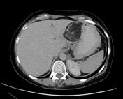
Image 1 CT (non-contrast) ( update )
Caption
was changed:
Case 1 -: hepatic angiomyolipoma
Image 2 CT (C+ portal venous phase) ( update )

Caption
was changed:
Case 2 -: focal hepatic steatosis
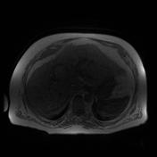
Image 3 MRI (T1 in-phase) ( update )
Caption
was changed:
Case 3 -: HCC in phase
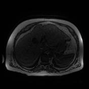
Image 4 MRI (T1 out-of-phase) ( update )
Caption
was changed:
Case 3 -: HCC out of phase