
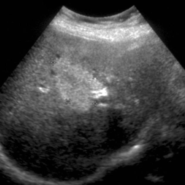
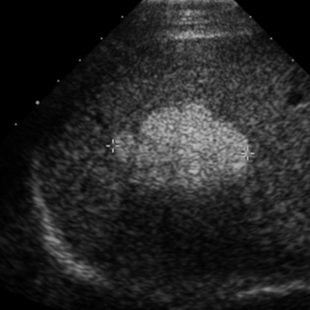
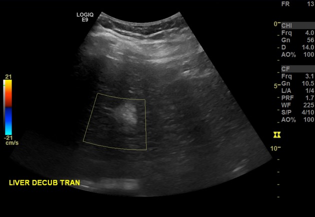
A hyperechoic liver lesion also known as an echogenic liver lesion, on ultrasound can arise from a number of entities, both benign and malignant. A benign hepatic hemangioma is the most common entity encountered, but in patients with atypical findings or risk for malignancy, other entities must be considered.
Benign
hepatic hemangioma: commonest hyperechoic liver lesion by far (present in 4% of the population) 1,4
hepatic adenoma with high fat content
-
focal fatty change: focal hepatic steatosis
rarely: multifocal hepatic steatosis
Malignant
-
colorectal carcinoma (up to 50% of hyperechoic liver metastases)
treated breast cancer
some sarcomas
hepatocellular carcinoma: particularly in a cirrhotic liver 3
The presence of hyperechogenicity can be a result of fat within a liver lesion 2, although some non-fat-containing lesions may also be echogenic (e.g. hepatic hemangioma).
On this page:
Radiographic features
Ultrasound
hypoechoic halo sign: considered a feature suggestive of malignancy
Some suggest pulse inversion harmonic imaging with quantitative evaluation as being useful in facilitating the differential diagnosis of hyperechoic focal liver lesions, where a lesion-liver ratio of ≥1 being predictive of a benign nature, assuming that malignant lesions show a ratio of <1 1.
Practical points
If a single, well-defined, homogeneous, echogenic mass is found in an asymptomatic patient, without a history of malignancy and without risk factors for liver tumors, then a diagnosis of hemangioma can be made on ultrasound without the need for another test 5. If an appropriate clinical history is not available, then a wider differential is appropriate. A "typical hemangioma" has been described as <3 cm 6, although this distinction appears to have originally meant to distinguish between "typical" and "cavernous" hemangiomas.


 Unable to process the form. Check for errors and try again.