
Liposarcomas are malignant tumors of fatty tissue and are the malignant counterpart to a benign lipoma. They are the second most common soft tissue sarcoma, after undifferentiated pleomorphic sarcoma.
On this page:
Epidemiology
Liposarcomas are typically found in adults, typically between the ages of 40 and 60, and are exceedingly rare in children.
Clinical presentation
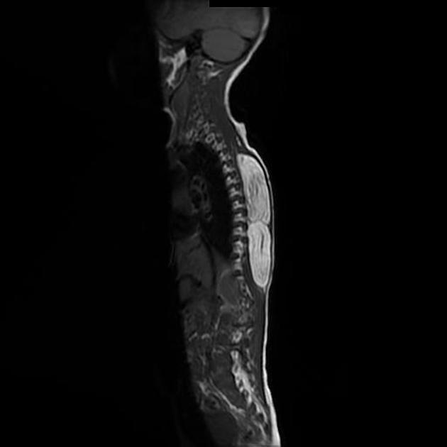
Symptoms vary by tumor location, usually a mass is described when involving the subcutaneous tissues and extremities, and a vague discomfort when intra-abdominal or intrathoracic.
Pathology
Thought to originate from mesenchymal cells, they are classified histologically into five types 1:
-
well differentiated liposarcoma
also known as atypical lipomatous tumor
most common (~50%) 4
has the highest amount of fat content
-
second most common
intermediate grade (myxoid) or high grade (round cell)
balanced chromosomal translocation t (12,16)
-
high grade, aggressive with frequent metastases
-
least common
high grade, aggressive with frequent metastases
-
mixed
Location
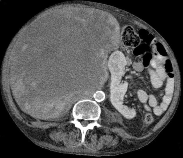
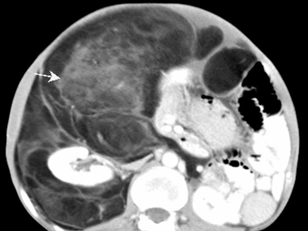
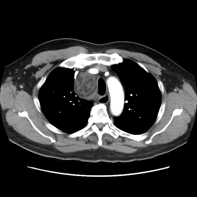
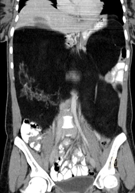
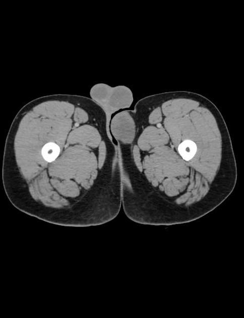
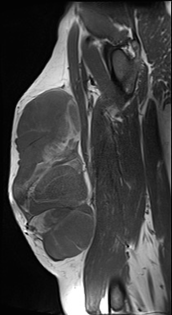
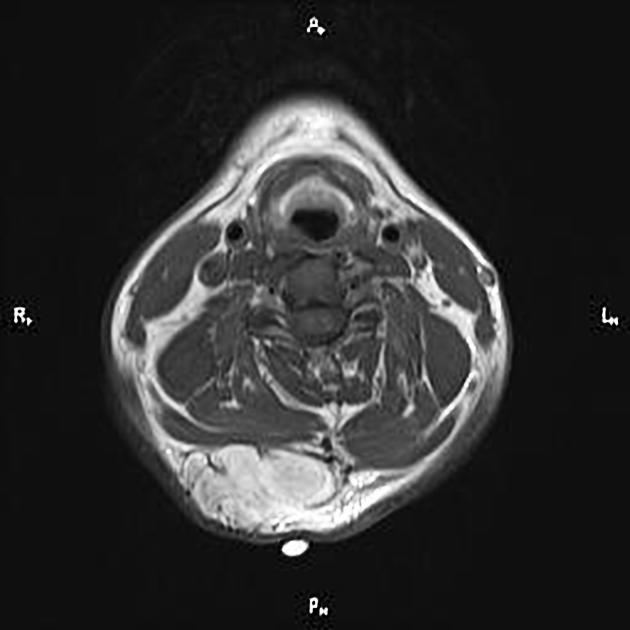
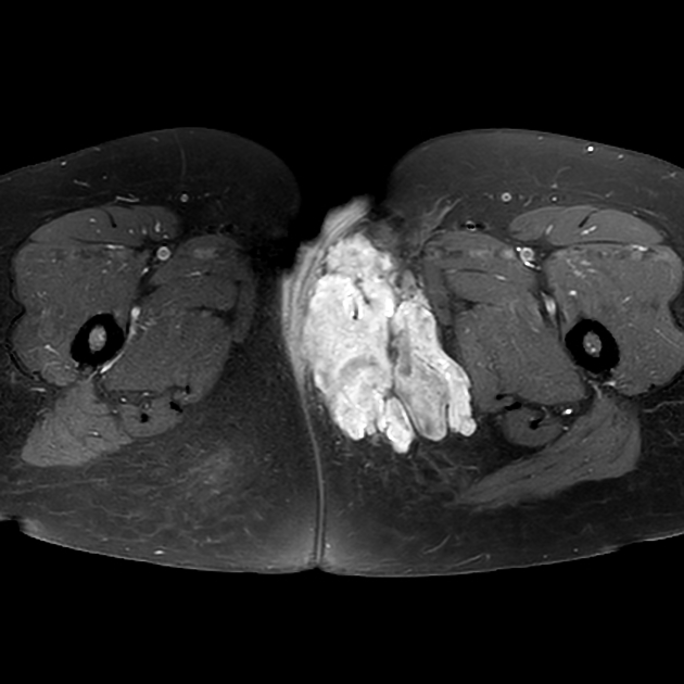
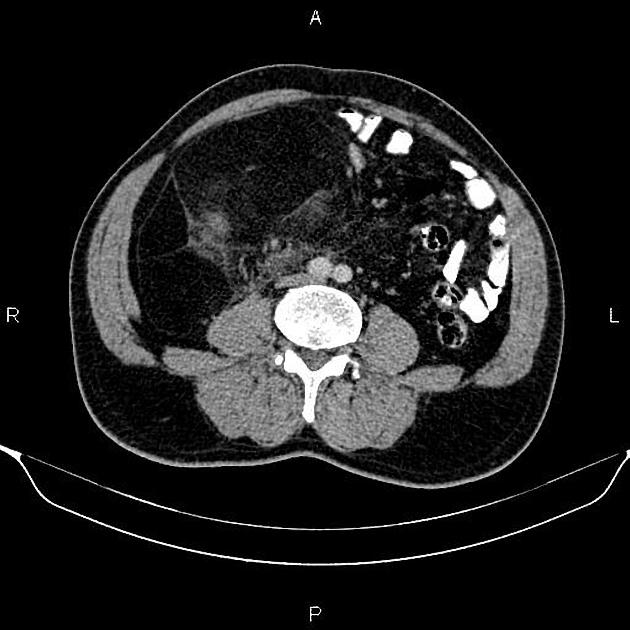
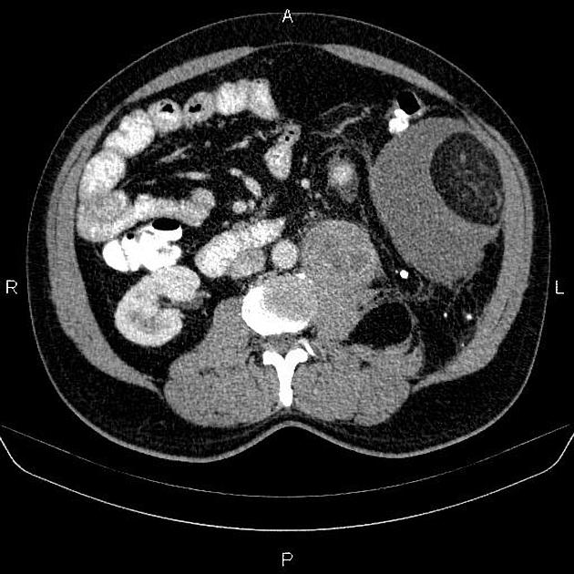
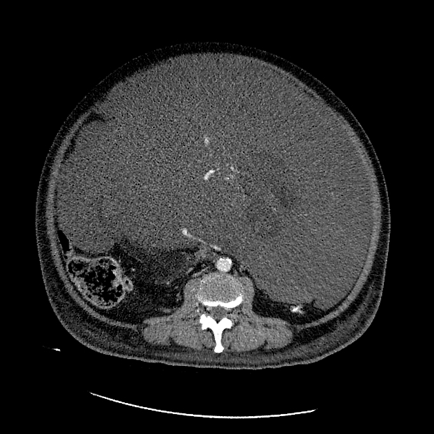
They are usually seen in the extremities (75%), most commonly the thigh, and are less commonly seen in the retroperitoneum, groin or elsewhere 2,9 (see: retroperitoneal liposarcoma).
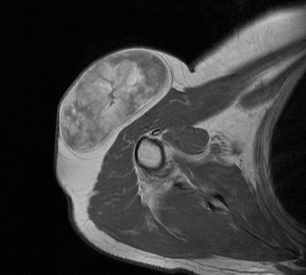
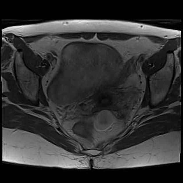
Radiographic features
CT
Liposarcomas have three CT patterns based on the amount and distribution of fat in the tumor:
solid: attenuation over +20 HU
mixed: areas of less than -20 HU and areas of over +20 HU
pseudocystic: homogeneous density between –20 and +20 HU
CT findings favoring a liposarcoma over a lipoma include:
inhomogeneous attenuation, with evidence of significant amounts of soft-tissue within the fatty mass
poor definition of adjacent structures
evidence of infiltration or invasion of mediastinal structures
calcification 8
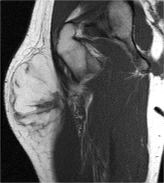
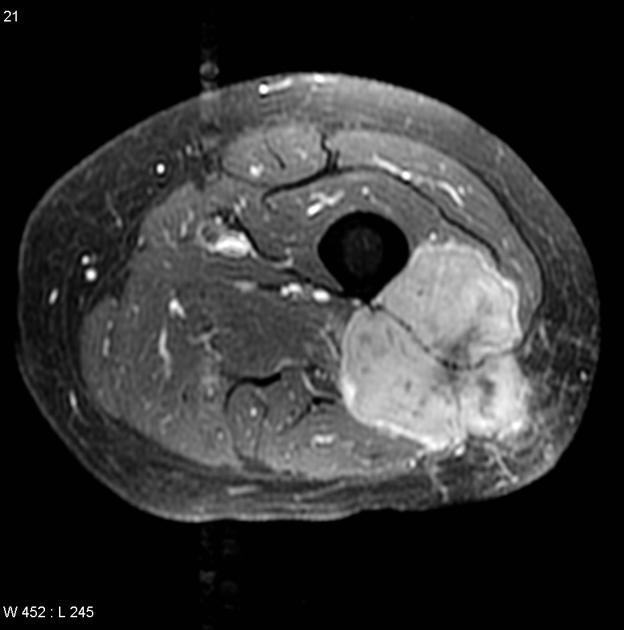
MRI
MRI appearance, as with CT, varies depending on the grade and amount of fatty tissue.
Low-grade lesions (atypical lipomas) are almost entirely fat signal with however thick septa, enhancement or evidence of local invasion. These features are used to distinguish these from simple lipomas 2.
The higher grade lesions are often devoid of macroscopic fat and have appearances similar to other sarcomas.
Treatment and prognosis
Both treatment and prognosis vary with location and grade.
Extremity liposarcomas, especially when well differentiated (atypical lipoma or atypical intramuscular lipoma) are indolent but nonetheless have a tendency to locally recur (0-69% of the time 2). The rate of recurrence is higher for deep lesions compared to superficial ones.
High-grade lesions, especially those of the retroperitoneum, have a poor prognosis, with a recurrence rate of ~75% (range 63-91%) 2.
Surgical treatment is with wide local excision, which accounts at least in part for the more favorable outcome for extremity lesions.
Differential diagnosis
General imaging differential considerations include:
undifferentiated pleomorphic sarcoma (UPS), formerly called malignant fibrous histiocytoma (MFH)
other sarcomas, e.g. fibrosarcoma, leiomyosarcoma
-
other fat-containing masses in the retroperitoneum


 Unable to process the form. Check for errors and try again.