
Fetal enteric duplication cysts are enteric duplication cysts presenting in utero.
On this page:
Epidemiology
Associations
They are associated with additional abnormalities in up to a third of the cases. These are most commonly vertebral anomalies 3.
Pathology
They result from an abnormal recanalization of the gastrointestinal tract. They comprise of a two-layer smooth muscle wall and an internal epithelium of a respiratory or intestinal type. These cysts may or may not communicate with the lumen of the gastrointestinal tract. They can be cystic or tubular (with the latter more likely to communicate with the gastrointestinal tract).
Location
They can occur anywhere along the gastrointestinal tract although a greater predilection towards the ileal region (~40%) 3.
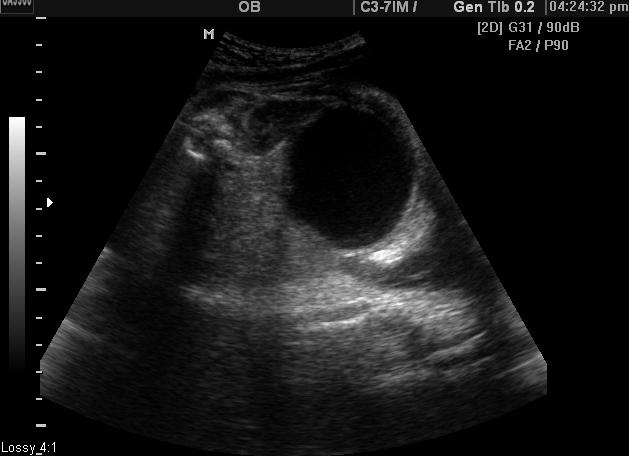
Radiographic features
Ultrasound
Usually seen as an anechoic cystic lesion within the abdomen that is separate from normal hollow structures such as the bladder and stomach.
Relatively characteristic signs that have been described include 1:
double wall sign: may not always be present
gut signature sign: may not always be present
Occasionally they can also present as an echogenic mass 3 (probably due to hemorrhage).
If there is a complicating bowel obstruction, there may be evidence of polyhydramnios.
Treatment and prognosis
Recognized complications include:
in utero bowel obstruction, sometimes with associated perforation
Differential diagnosis
Considerations on ultrasound for an anechoic mass include:


 Unable to process the form. Check for errors and try again.