
Hemorrhagic pulmonary metastases are those which tend to be complicated by pulmonary hemorrhage within them, resulting in characteristic imaging appearances. Metastases of some tumor histologies are more likely to hemorrhage -- knowledge of this can help refine the differential diagnoses.
On this page:
Pathology
Hemorrhage is mostly thought to be due to fragility of neovascular tissue that leads to a rupture of the vessel.
Recognized types generally tend to have high vascularity within them and include 1,2
- angiosarcoma - metastatic angiosarcoma to the lungs
- choriocarcinoma - metastatic choriocarcinoma to the lungs
- renal cell carcinoma
- melanoma
Radiographic features
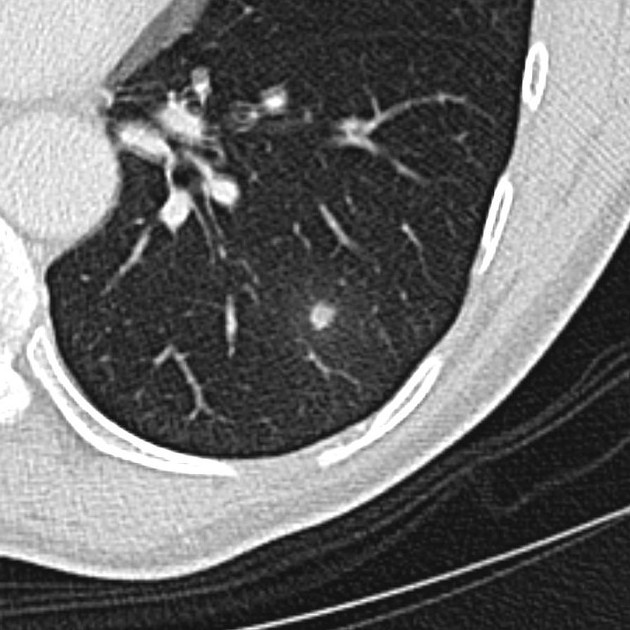
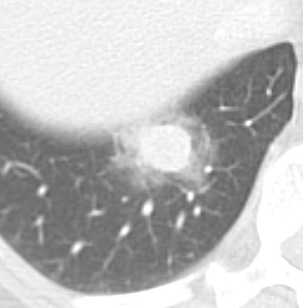
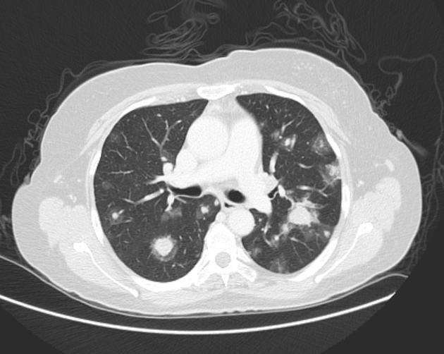
CT chest
If there is a peri-tumoral hemorrhage this may be seen as a nodule or mass which is surrounded by a halo of ground-glass opacity (CT halo sign) or an ill-defined fuzzed-out margin.
Differential diagnosis
For a suspected nodule or mass giving a CT halo sign - see differential for a CT halo sign.


 Unable to process the form. Check for errors and try again.