
The hyoid bone is a midline "U or horseshoe-shaped" bone that serves as a structural anchor in the mid-neck. It is the only bone in the human body that does not directly articulate with another bone (other than sesamoids). It is a place of convergence of multiple small neck muscles that permit the pharyngeal phase of swallowing. The location of structures in the neck is often described in terms of relation to the hyoid (i.e. suprahyoid neck; infrahyoid neck).
On this page:
Summary
-
location: midline structure located between the mandible and the thyroid cartilage and anterior to the epiglottis at the level of C3/C4
the level of the hyoid separates lymph node levels II from III in the lateral neck and levels Ia and VI in the anterior neck
articulations: none
blood supply: branches of the external carotid artery
relations: numerous muscles insert on the hyoid, detailed below
Gross anatomy
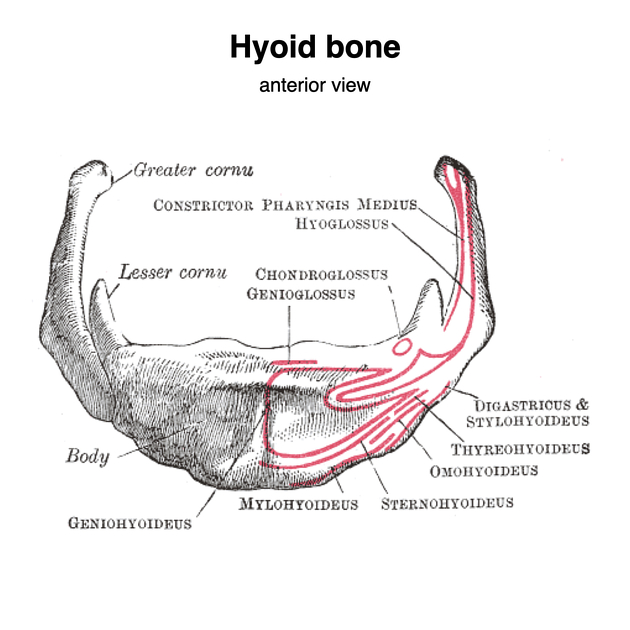
The hyoid bone is located in the mid-neck, above the thyroid cartilage, anterior to the pharynx. It is divided into 5 parts:
body with a transverse ridge running along its superior anterior surface and an incomplete vertical ridge in the midline on its superior anterior surface
-
two greater horns or cornua which are long slender and slightly curved projections that project posteriorly
the posterior end of the greater horn forms a small tubercle
the joint between the greater horn and the hyoid body is cartilaginous and usually ossifies by the 3rd decade
-
two lesser horns or cornua which are short conical projection that project superiorly from the junction of the greater horn and body
the joint between the lesser horn and the hyoid body is fibrous
occasionally a synovial joint exists between the lesser and greater horns that can ossify
Attachments
Multiple small neck muscles insert onto different parts of the hyoid 4:
-
body
-
digastric muscles (indirectly)
stylohyoid muscle (indirectly)
genioglossus muscle inferior fibers (indirectly)
-
-
body and greater horn
-
greater horn
Several ligaments also attach to the hyoid:
-
lesser horn
-
body
-
body and greater horn
thyrohyoid membrane, including median thyrohyoid ligament
Blood supply
Branches of the external carotid artery, predominantly the infrahyoid branch of the superior thyroid artery, and branches of the lingual artery.
Relations
The position of the hyoid defines a few lymph node levels of the neck:
anteriorly: hyoid separates levels Ia (above) from VI (below)
laterally: hyoid separates level II from level III
Variant anatomy
asymmetry (common)
long slender lesser horns due to fusion with the ossified stylohyoid ligament
failure of fusion of the greater horns to the body
absent lesser horns
inferiorly positioned anterior to the thyroid cartilage
pierced by the thyroglossal duct
lingula - midline superior projection from the body
absent (rare)
Radiographic features
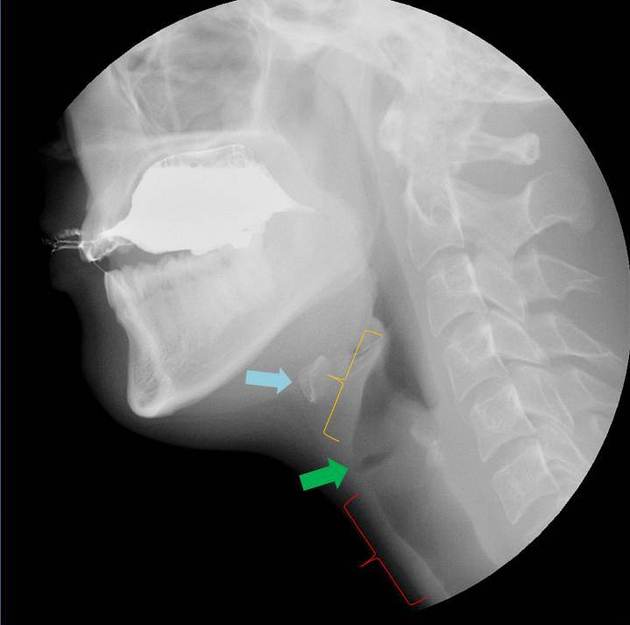
The hyoid is the ossified horseshoe-shaped structure just superior to the thyroid cartilage.
Related pathology
hyoid bone fracture which has a high association with strangulation
movement of the hyoid (hyoid elevation) is an indication of the strength of the pharyngeal muscles during a modified barium swallow study ref


 Unable to process the form. Check for errors and try again.