
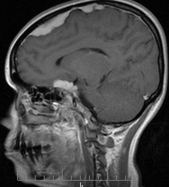
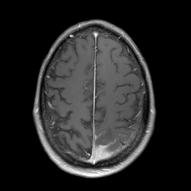
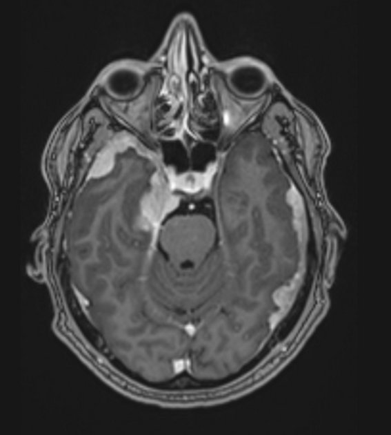
Hypertrophic pachymeningitis is a condition where there is localized or diffuse inflammatory thickening of the dura. On imaging, it presents as a localized, multiple, or diffuse enhancing dural thickening commonly forming mass-like lesions.
On this page:
Clinical presentation
The clinical presentation may be varied 7. Common clinical features include headache and cranial nerve palsies 7.
Pathology
Etiology
It can result from a number of causes which include 5-8:
idiopathic
-
infective
-
inflammatory 9
-
other
hemodialysis
Radiographic features
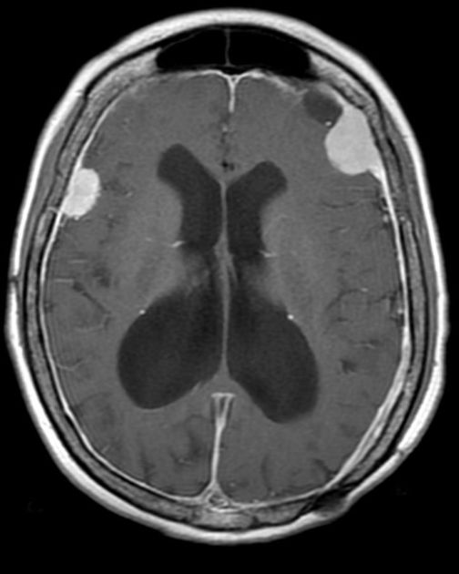
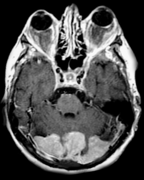
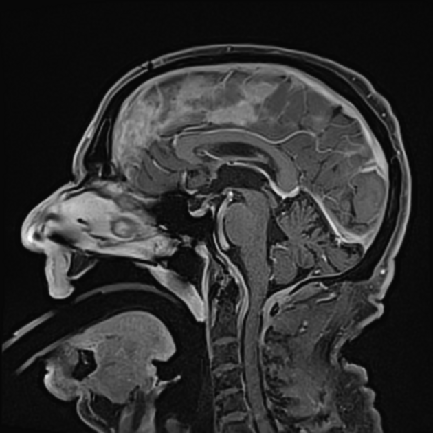
MRI
localized, or less often, diffuse dural thickening
may uncommonly depict mass-like thickening, termed tumefactive hypertrophic pachymeningitis 1
Signal characteristics:
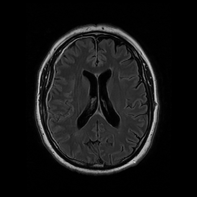
T1: thickened areas are hypointense to brain parenchyma 6
T1 C+ (Gd): dural enhancement
T2: thickened areas are hypointense to brain parenchyma 6
Treatment and prognosis
Management depends on the underlying cause, and includes immunosuppression in idiopathic cases 7.
Differential diagnosis
General imaging differential considerations include:


 Unable to process the form. Check for errors and try again.