
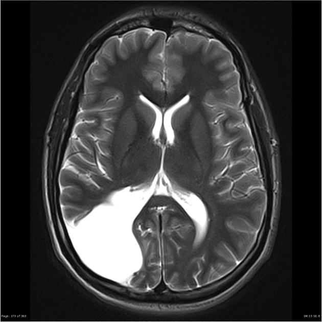
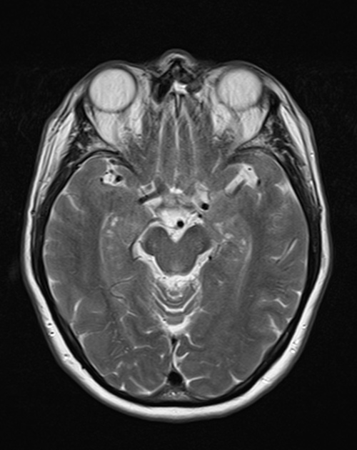
Intracranial non-neoplastic cysts are common findings on MRI and CT brain scans.
On this page:
Location-based diagnostic approach
A location-based approach is useful in establishing an appropriate diagnosis; some locations are virtually pathognomonic for certain lesions e.g. colloid cyst.
Many cysts may occur in more than one location (midline or off-midline) e.g. arachnoid and epidermoid cysts.
Features which help the diagnostic approach for intracranial cysts:
- is the cyst extra-axial or intra-axial cyst?
- is it supra- or infratentorial?
- if it is extra-axial, is it midline or off-midline?
- if it is intra-axial, is it parenchymal or intraventricular?
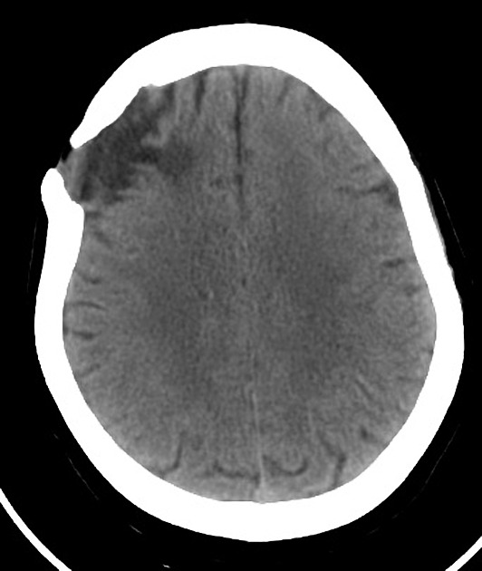
Extra-axial cysts
Supratentorial
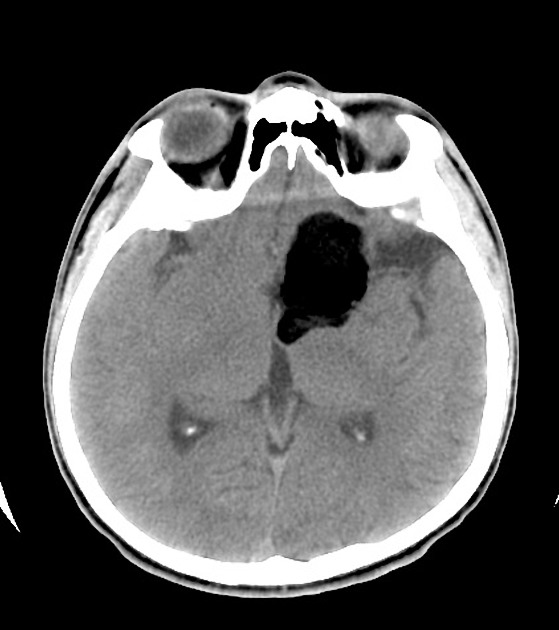
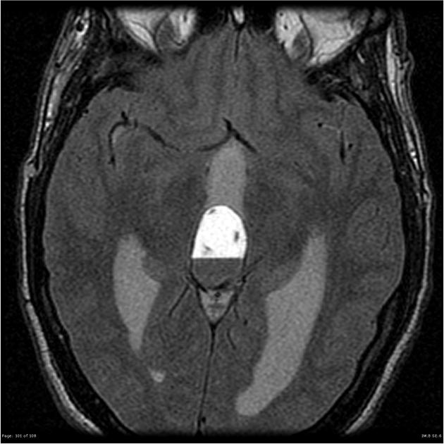
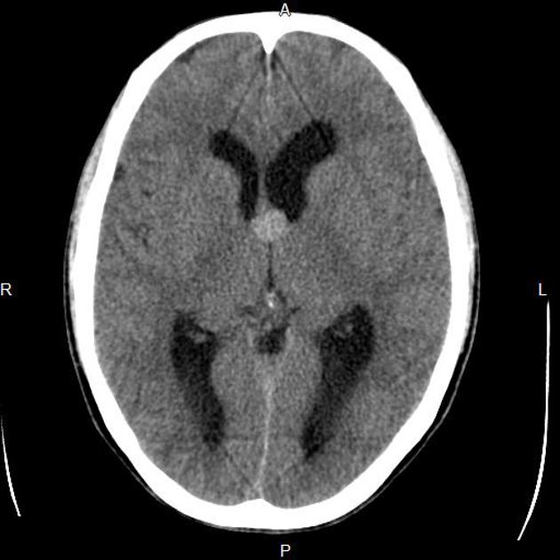
Midline
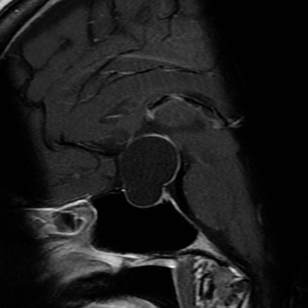
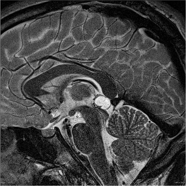
- Rathke cleft cyst
- arachnoid cysts: at suprasellar cistern
- dermoid cyst
- pineal cysts
Off-midline
- arachnoid cysts
- epidermoid cysts
- leptomeningeal cyst
- non-neoplastic tumor-associated cysts (TACs)
- choroid fissure cyst
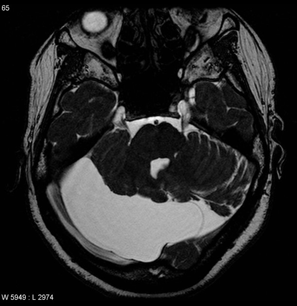
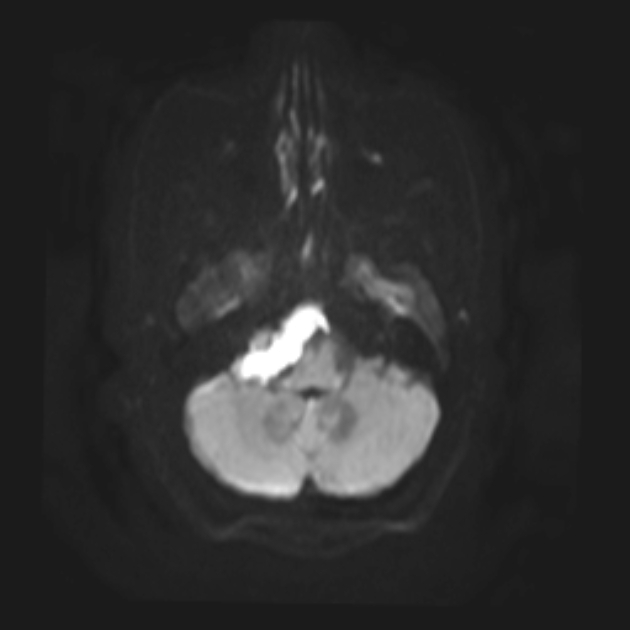
Infratentorial
Midline
- neuroenteric cyst
- arachnoid cysts: usually in the midline cisterna magna
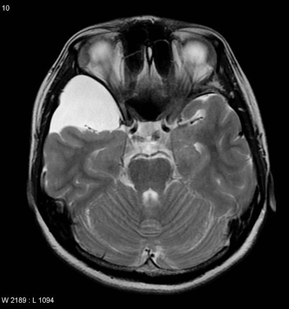
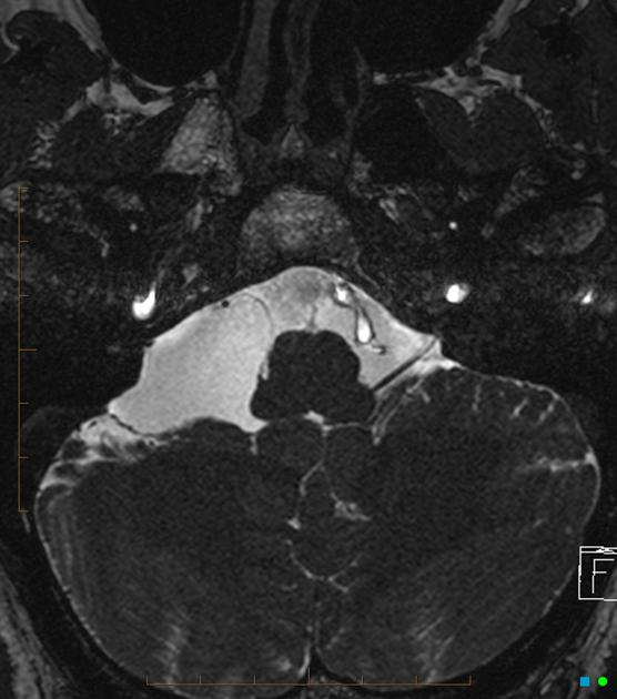
Off-midline (usually occurs in the cerebellopontine angle (CPA))
Intra-axial cysts
Intraventricular cysts
Supratentorial
Infratentorial
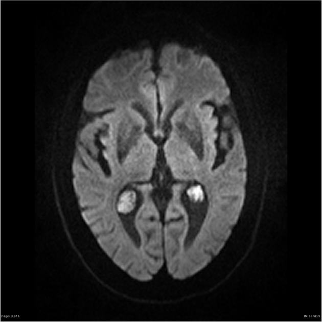
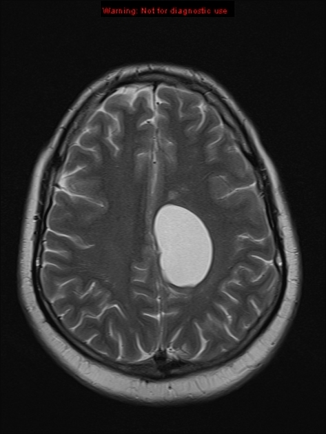
Intra-parenchymal
Supratentorial
Infratentorial
- enlarged perivascular spaces (dentate nuclei)
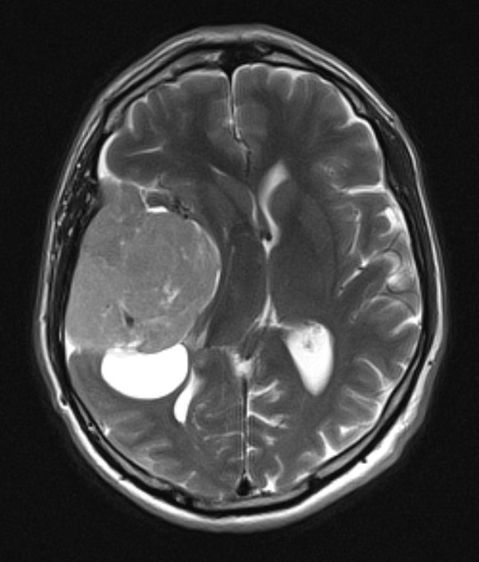
Differential diagnosis
- parasitic cysts
- cystic tumors
- cystic malformation: Dandy Walker malformation
- scalp and skull cysts e.g. sebaceous cysts


 Unable to process the form. Check for errors and try again.