
The lumbar spine (often shortened to L-spine) consists of five adjacent vertebrae of the lower vertebral column, in some cases it is possible to find 4 or 6 vertebrae as an anatomical variant. They participate in the lumbar lordosis, a natural curve in the spine, that is convex anteriorly.
Articulations of the facet (zygapophyseal) joints permit flexion/extension and abduction movements. Rotation is greatly limited, and may occur only slightly at the lumbosacral joints.
For a basic description of the anatomy of a generic vertebra, see vertebrae.
On this page:
Gross anatomy
Similar to other vertebrae, the osteology of each lumbar vertebrae can be subdivided anteriorly to posteriorly:
body: kidney shaped, convex anteriorly, flattened posteriorly; resulting vertebral canal is triangular in cross-section
pedicles: project directly posteriorly, attached to the upper half of the body
transverse processes: spatulate, project laterally on both sides; L3 most often has the longest transverse processes of the lumbar spine, a fact that can be used to number the vertebrae
lamina: broad, thick, overlap minimally
articular processes (superior and inferior): lie at the lateral angle of the laminae (junction with pedicle), connected via the pars interarticularis
spinous process: single, short, thick, roughly horizontal, hatchet-shaped (upper border is straight, lower border curves down)
L1 to L4 are considered typical lumbar vertebrae, whereas due to various and distinctive differences the fifth lumbar vertebra is considered atypical.
Articulations
Vertebrae articulate with one another via:
intervertebral discs (superior and inferior): wedge shaped (taller anterior, shorter posterior), and contribute most to the lumbar lordosis
-
facet (zygapophyseal) joints: obliquely orientated, cylindrically-shaped articular surfaces
superior articular facets are concave and face posteromedially
inferior articular facets are convex, and face anteriorly
Inferior articular process of the vertebrae above always lies posterior to the superior articular process of the vertebrae below.
Attachments
A large number of attachments occur at the transverse process. On its anterior surface, a vertical ridge serves as an important landmark.
medial to ridge: psoas muscle
at the ridge: psoas fascia, anterior layer of the lumbar fascia, medial and lateral arcuate ligaments (at L1)
lateral to ridge: quadratus lumborum partially inserts
transverse process tip: middle layer of lumbar fascia
Posterior surface receives attachments of erector spinae. Back muscles (multifidus, longissimus) attach to the mammillary process and accessory tubercle:
mammillary process: located on the superior articular process, behind the margin of the articular facet, projects posteriorly
accessory tubercle: located at the root of the transverse process, projects posteriorly
Relations and/or boundaries
Can be superimposed onto the three-column concept.
Anterior column
anterior longitudinal ligament (ALL): anterior to vertebral body
Middle column
posterior longitudinal ligament (PLL): lies posterior to vertebral body
-
centrally
basivertebral veins and internal vertebral venous plexus
-
laterally
lumbar fascia
intertransverse ligaments
regional/segmental lumbar arteries and veins
Posterior column
external vertebral venous plexus (lying within muscle)
Arterial supply
Arterial supply comes from regional/segmental lumbar arteries.
Venous drainage
Venous drainage is complex. Lumbar vertebral bodies are posteriorly perforated by a pair of basivertebral veins that drain into the internal vertebral venous plexus. Regional/segmental lumbar veins also contribute.
Variant anatomy
transitional vertebrae of the lumbar spine are possible at multiple levels in the lumbar spine
limbus vertebrae represent herniation of the disc nucleus pulposus through the superior endplate and can simulate a fracture
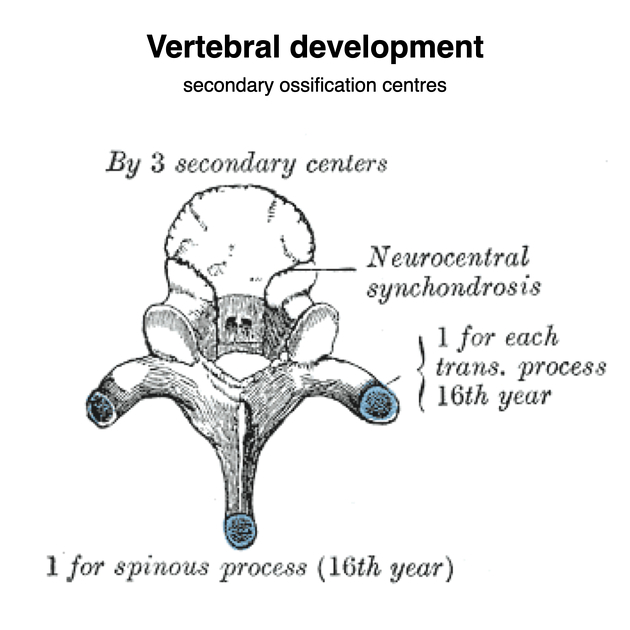
Development
Ossification centres of generic vertebrae are covered here. Transverse processes are actually fused ribs. Mammillary process and the accessory tubercle are the remnants of the true transverse process, typically seen on thoracic vertebrae.


 Unable to process the form. Check for errors and try again.