
Mesoblastic nephroma, also sometimes known as a congenital mesoblastic nephroma (CMN), Boland's tumor or fetal renal hamartoma, is generally a benign renal tumor that typically occurs in utero or in infancy.
On this page:
Epidemiology
It is the most common neonatal renal tumor. Diagnosis is usually made in the antenatal period (16% of cases) or soon after birth 14. The tumor accounts for ~5% of renal neoplasms in children 3,7. The median age of diagnosis is 3 months of age, and 90% are diagnosed by the age of 1 year 11,14. There is a slight male predilection (male to female ratio 1.5:1) 14.
Associations
Clinical presentation
The most common clinical presentation is a palpable abdominal mass, with hematuria and hypertension occurring less frequently 14.
Pathology
It is a mesenchymal tumor, which macroscopically is a solid un-encapsulated mass often occurring near the renal hilum. It tends to invade the surrounding structures and renal parenchyma. Hemorrhage and necrosis are infrequent. Histologically, it is typically composed of connective tissue growing between nephrons, usually replacing most of the renal parenchyma.
The classic cytological description of the lesion is that of cellular clusters of spindle cells, mild nuclear pleomorphism, mitotic activity and no blastema.
Subtypes
There are two main pathological variants:
classic: accounts for less than one third of cases of CMN 11
-
more heterogeneous in appearance on imaging
tends to be larger and presents later in infancy (>3 months of life 11)
may exhibit aggressive behavior including vascular encasement and metastasis 5
Radiographic features
Plain radiograph
Non-specific and not an imaging modality of choice, but may demonstrate a soft tissue mass displacing bowel. Calcification is rare 3.
Ultrasound
Sonographic appearance varies with the pathological variant 6. In general it is a well-defined mass with low-level homogeneous echoes. The presence of concentric echogenic and hypoechoic rings can be a helpful diagnostic feature in the classic subtype, but may also be seen in the cellular subtype 11. A more complex pattern due to hemorrhage, cyst formation and necrosis favors the cellular variant. Color Doppler may show increased vascularity. Uncommonly the tumor may appear predominantly cystic 11.
Antenatal ultrasound may also show associated polyhydramnios 14.
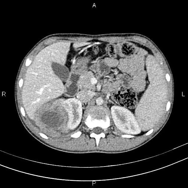
CT
Solid hypoattenuating renal lesion with variable contrast enhancement. Cystic areas, necrosis, and hemorrhage are uncommon (only in cellular type) 5. Typically no calcification is seen, but hyperdense foci from hemorrhage may be seen in the cellular subtype 13.
MRI
The best antenatal modality antenatally, which can be used to assess anatomic relationships.
Unless complicated by necrosis and hemorrhage (which is uncommon), general signal characteristics include:
T1: iso to hypointense 8; hyperintense foci may relate to hemorrhage in the cellular subtype 13
T2: variable, from markedly hypointense to hyperintense 11
DWI/ADC: the solid portion of the tumor may diffusion restrict, likely related to increase cellularity 12
Treatment and prognosis
The majority are benign tumors with an excellent prognosis if completely excised 15. The cellular variant can be more aggressive and is associated with worse outcomes 15.
Complications
Potential complications with large tumors include 11:
preterm delivery
abdominal dystocia at birth
arterio-venous shunting with subsequent development of hydrops fetalis
Differential diagnosis
Wilms tumor: older age group (typically after 1 year of life, with peak incidence between 3 and 4 years of life)


 Unable to process the form. Check for errors and try again.