
Mesothelioma is an aggressive malignant tumor of mesothelium and 90% of tumors arise from the pleura.
This article is about the pleural form of the disease, other locations include 17:
peritoneal mesothelioma (~10%)
pericardial mesothelioma (<1%)
On this page:
Epidemiology
Mesothelioma is an uncommon entity and accounts for ~15% (range 5-28%) of all malignancies that involve the pleura 1,7. There is a strong association with exposure to asbestos fibers with a 10% lifetime risk; 40-80% of affected patients have a known history of asbestos exposure 1 and risk is proportionate to duration and quantity 20. Para-occupational exposure of household members of asbestos-exposed workers is documented 14,20.
Crocidolite is the main causative fiber type. Sources of asbestos exposure are predominantly mining, construction, lagging and machinery mechanics and 60-80% of cases occur in males, typically with a latent period of 20 to 35 years following exposure 1,5,6. Regional hotspots are related to local industries e.g. the historic shipbuilding industry in Belfast, Northern Ireland.
There is no convincing evidence for an association with smoking 6.
Clinical presentation
Typically patients present with dyspnea and low back non-pleuritic chest pain. Pleural effusions are seen in the vast majority of patients at some stage during their disease 20. Up to 25% of patients have metastatic disease at the time of presentation if staged with FDG PET 5.
Typically mesothelioma is a locally-aggressive disease and distant extranodal metastases are uncommon in life15. In a postmortem study of 318 patients, 55% patients were found to have extrathoracic metastases, the commonest sites being the liver (32%), spleen (11%), thyroid (7%) and brain (3%) 16.
Pathology
Etiology
asbestos fiber exposure: causes the majority of cases
erionite - a fibrous zeolite used in the building sector (particularly in Turkey) 12 and for road-surfacing. Inhalational exposure occurs during mining, production and use and the mineral is highly toxic 24. Exposure correlates with increased risk of lung cancer and mesothelioma 25-28. Erionite has been included by the International Agency for Research on Cancer (IARC), as a human carcinogens (group 1 - volume publication year 2012) 29.
oncogenic simian virus 40 (SV40), belonging to the Polyoma viruses 13,22. The transforming and oncogenic potential of the SV40 virus resides mainly in the viral oncoprotein large T antigen 23.
radiation exposure 13
Histology
There are three histological types of mesothelioma:
epithelial: ~60%
mixed: 25%
sarcomatoid: 15%
The cytological and histological diagnosis can be difficult, with mesothelial hyperplasia and metastatic adenocarcinoma appearing similar. Specific markers are helpful including:
calretinin
epithelial membrane antigen
cytokeratin
mesothelin (elevated in 84% of malignant mesothelioma versus <2% with other pleural diseases 6)
Subtypes such as multicystic/cystic mesothelioma are rarer and less aggressive.
Radiographic features
See: staging of malignant pleural mesothelioma.
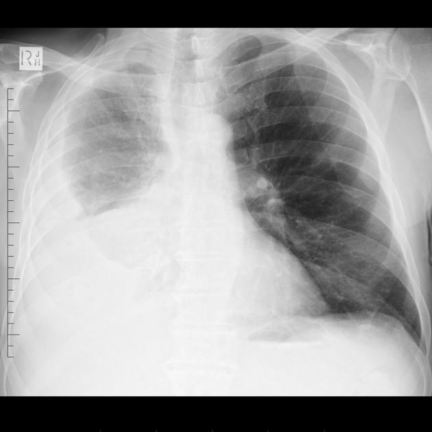
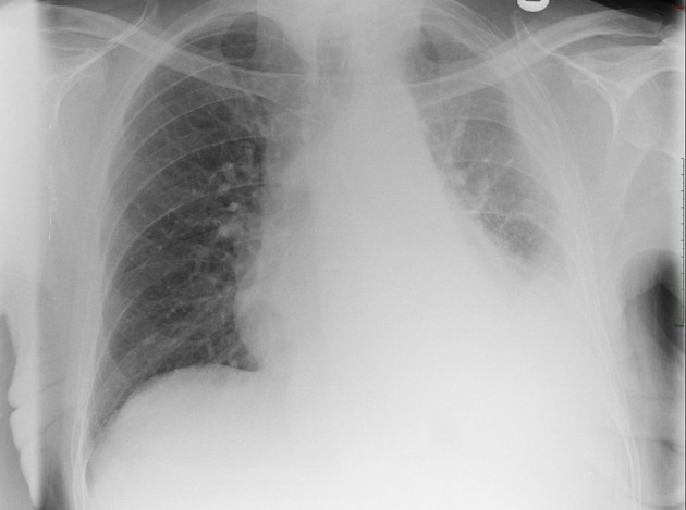
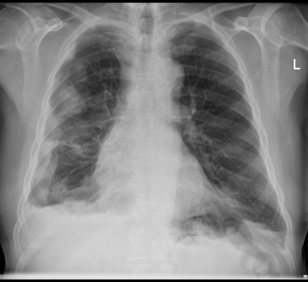
Plain radiograph
Chest radiographs are non-specific and of limited utility 6. The following features may be evident:
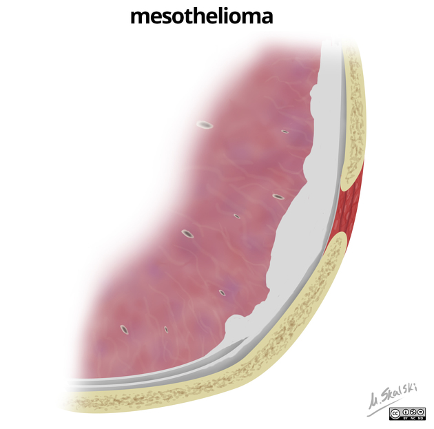
pleural opacity which may extend around and encase the lung
reduced volume of the affected hemithorax, resulting in ipsilateral shift of the mediastinum (common) 4
rib destruction or extension beyond the lateral and anterior margins of the chest wall
+/- pleural effusion; most commonly is unilateral and exudative or hemorrhagic in nature, with frozen hemithorax (not causing mediastinal shift)
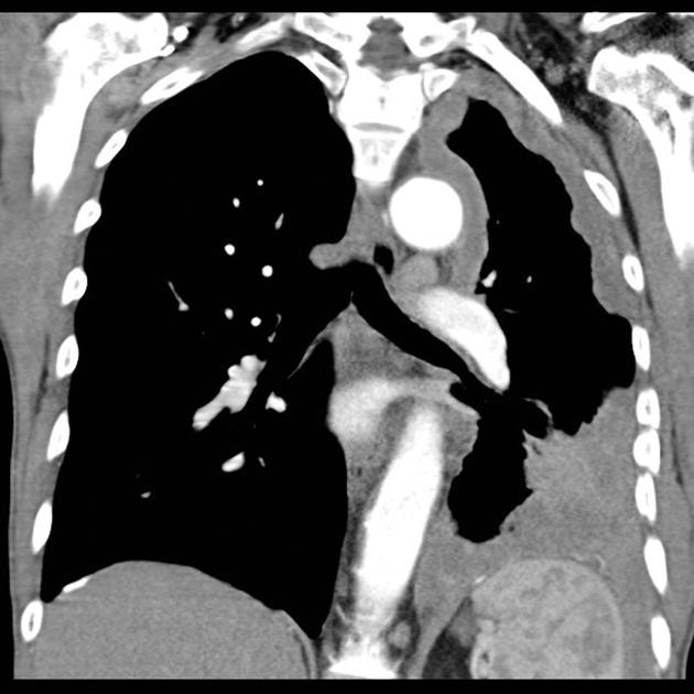
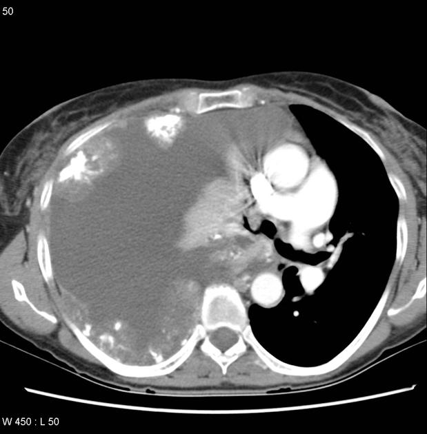
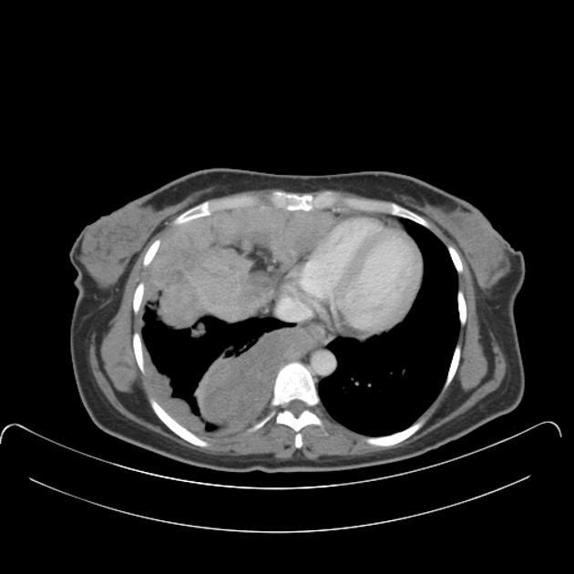
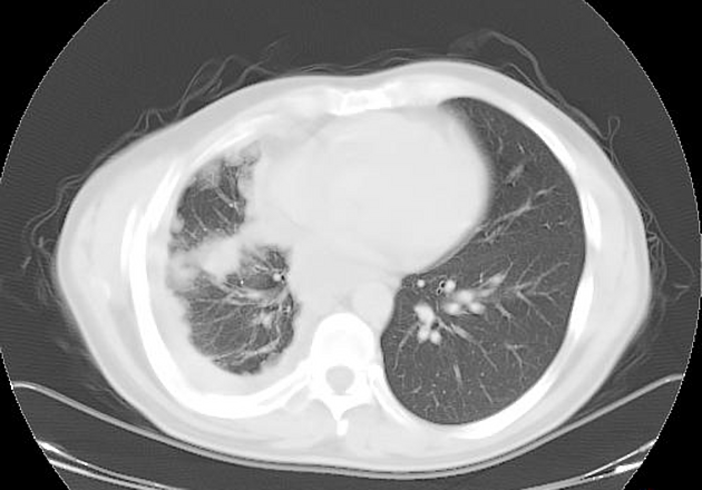
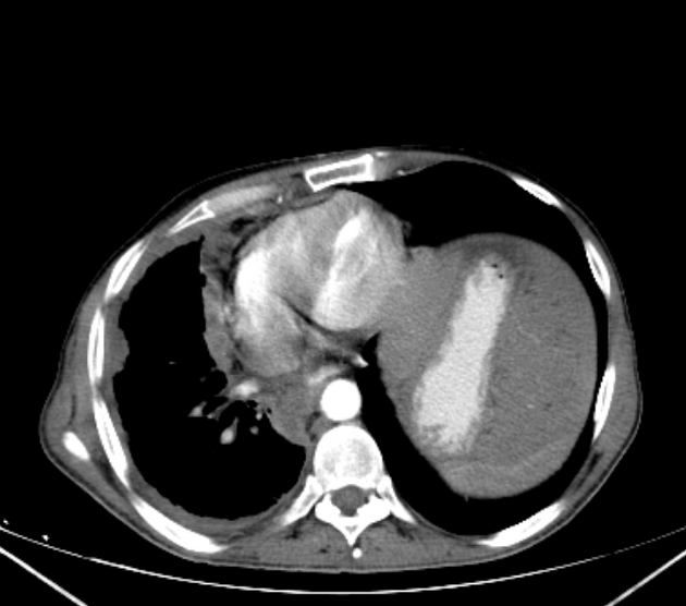
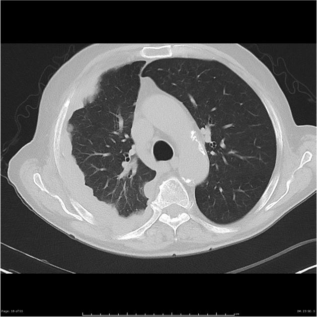
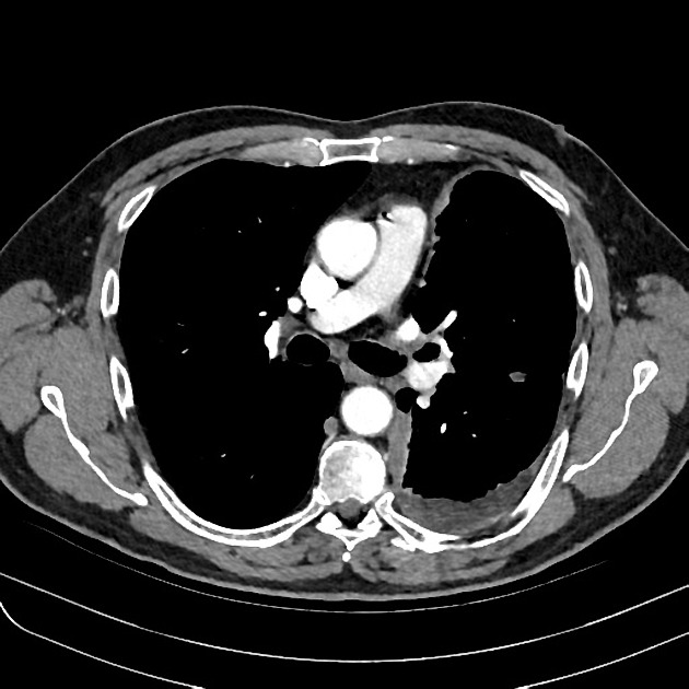
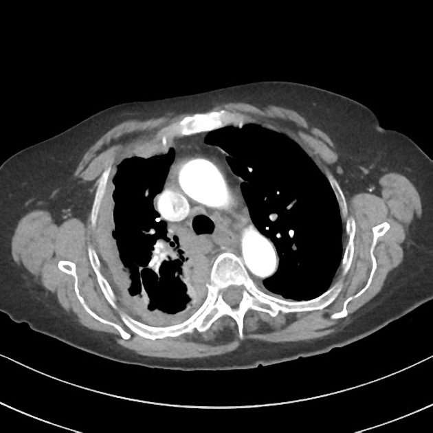
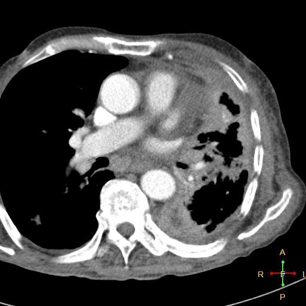
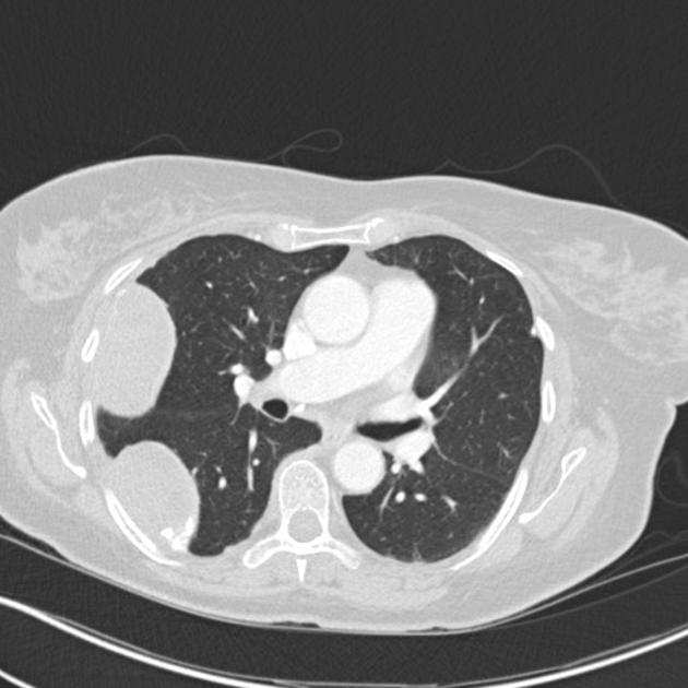
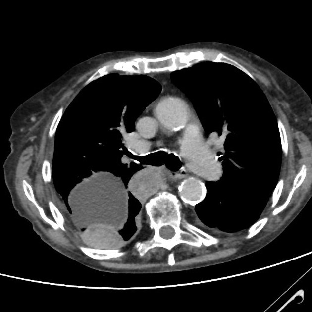
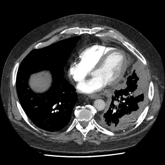
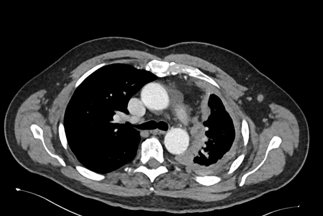
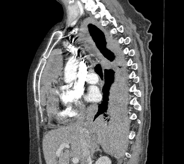
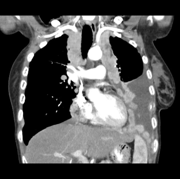
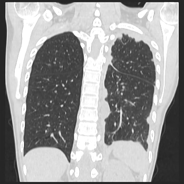
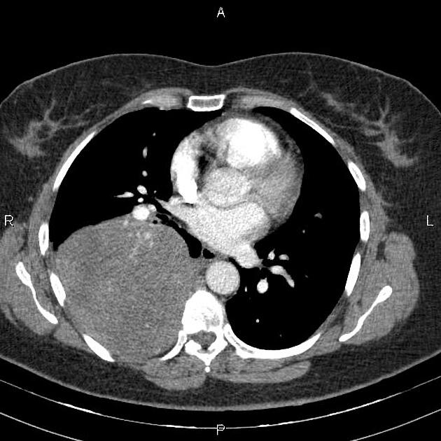
CT
CT is most commonly used for imaging assessment of mesothelioma, and sufficient for accurate staging of disease in most patients.
-
pleural mass or nodular thickening of soft tissue attenuation
tends to cause "inward" contraction of the hemithorax, e.g. ipsilateral mediastinal shift
-
pattern of spread
-
pattern of spread initially to adjacent pleura
involvement pleural fissures
eventually grows toward lung encasement ("pleural rind')
-
predilection for local invasion
involvement of chest wall, diaphragm, and mediastinal content typical 1,2,4
-
chest wall involvement
infiltration of the extrapleural fat plane 4
obvious direct extension in bone or muscle 4
known to invade along prior catheter and biopsy tracks 18
pericardial effusion may represent transpericardial extension 3,4
frequent metastasis to local lymph nodes and contralateral lung
-
-
calcification
seen in 20% - usually represents engulfment of calcified pleural plaques rather than true tumor calcification 4
sarcomatoid variants may contain calcific osteosarcoma or chondrosarcomatous components
An uncommon variant is the solitary mediastinal malignant mesothelioma which has appearances reminiscent of a solitary fibrous tumor of the pleura 1.
MRI
MRI, although not routinely used, may have a role in refining the staging and better delineating the extent of the disease in surgical candidates especially concerning the chest wall and diaphragmatic invasion 4.
T1: iso to slightly hyperintense cf. muscle 4,6
T2: iso to hyperintense cf. muscle 4,6
T1 C+ (Gd): enhancement usually present
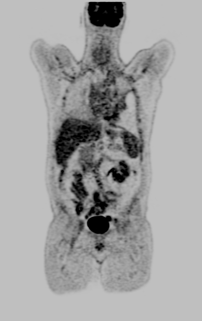
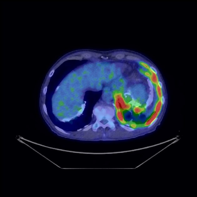
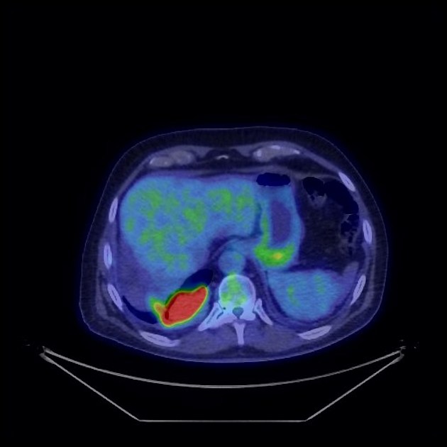
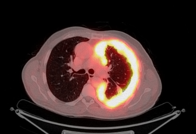
PET
Positron emission tomography is becoming useful in two clinical settings 4:
differentiating between benign and malignant asbestos-related pleural thickening
assessing for nodal metastases
In addition, there appears to be a correlation between the degree of FDG uptake and the biological aggressiveness of the tumor, which may help to guide treatment 4.
Treatment and prognosis
Treatment continues to be challenging and the long-term survival is poor. Single modality treatment (surgery, radiotherapy, chemotherapy, immunotherapy and even photodynamic therapy) have not been shown to improve survival 3. Multi-modality treatment has had some impact on favorable subgroups (early disease, and epithelioid histology). Treatment includes:
adjuvant chemotherapy
radiotherapy
The prognosis is poor for all tumor types with a median overall survival without treatment of 4-12 months 3. In favorable patient subgroups up to 45% 5-year survival may be achievable 3, however even with aggressive multi-modality therapy overall 5-year survival remains poor (3-18%) 3 with a median survival time of approximately 18 months 4.
Differential diagnosis
The differential is dependent on the exact nature of tumor involvement and the modality. General imaging differential considerations include
pleural effusion (especially if loculated): on radiographs
pleural metastases (especially with pleural carcinomatosis)
peripheral lung cancer
other pleural-based tumors
-
tumor-like conditions of the pleura
pleural fibrosis from infective/inflammatory source (e.g. actinomycetes, tuberculosis)
Practical points
some caution is raised with performing an image-guided biopsy, as mesothelioma can potentially cause tumor seeding along the biopsy track (with a reported incidence of around 4%) 21


 Unable to process the form. Check for errors and try again.