
Microcystic architecture is a rare meningioma variant which leads to atypical imaging appearances and thus can pose a diagnostic challenge. They should not be confused with cystic meningiomas, which represent a variant where large cysts are present within or adjacent to the tumor.
These tumors do not appear to differ epidemiologically or clinically from the more common subtypes. As such for a general discussion, please refer to the meningioma article.
On this page:
Pathology
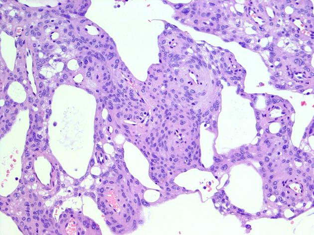
Microcystic meningiomas account for only 1.6% of intracranial meningiomas 1. The microcysts represent extracellular spaces, scattered throughout the meningioma substrate. These tumors usually also show abundant vascularity.
Radiographic features
On both CT and MRI, the imaging appearances are dominated by the high water content of these tumors.
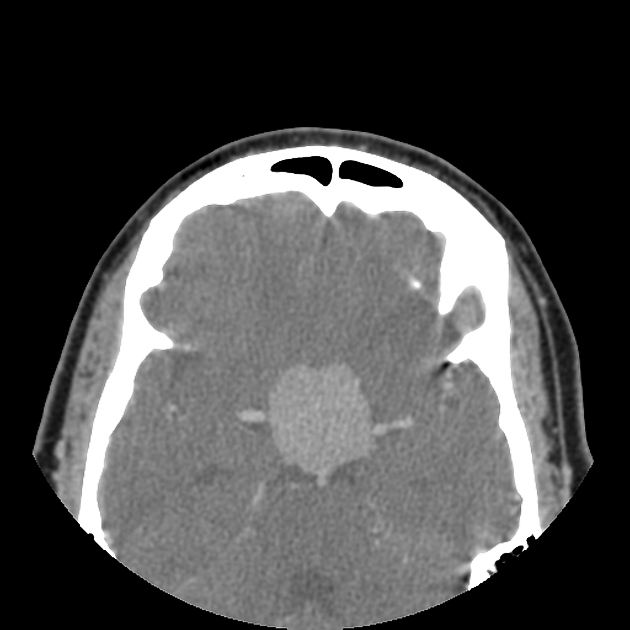
CT
Typically these lesions are of low density but usually still have intense homogeneous enhancement. Hyperostotic changes in the adjacent skull are seen in approximately half of cases 2.
MRI
-
T1
low intensity (similar to fluid)
often have some solid components which are of more intermediate signal intensity
-
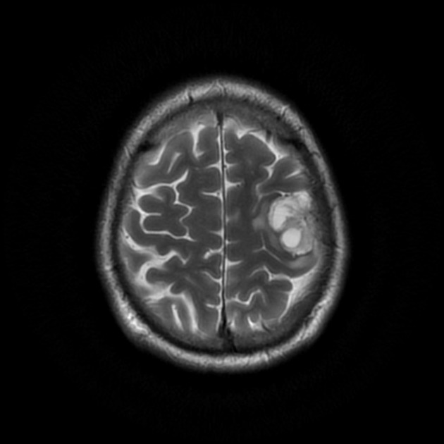
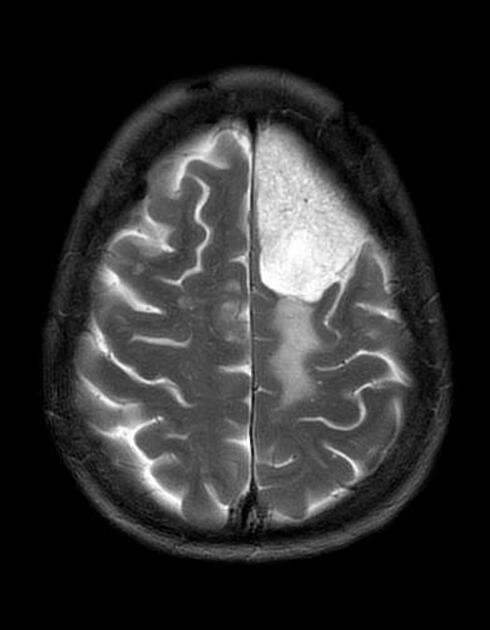
T2
very high signal
associated edema in the adjacent brain is common 2
-
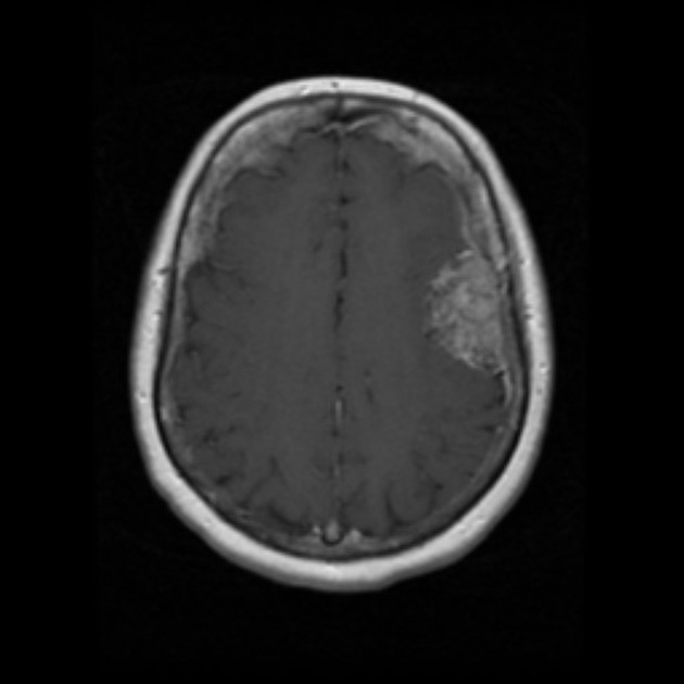
T1 C+ (Gd)
variable 1,2
usually intense homogeneous enhancement
some tumors enhance only partially or very little
DWI / ADC: facilitated diffusion
DSA
The degree of angiographic vascularity can often be predicted based on the degree of enhancement, with strong and uniform contrast enhancement demonstrating the main supply from both meningeal (external carotid artery / vertebral artery) and pial vessels (internal carotid artery / vertebral artery).
Differential diagnosis
In cases where enhancement is prominent, there is usually little differential, although it is probably worth considering exophytic or cortical tumors (e.g. pleomorphic xanthoastrocytoma (PXA)).
In tumors where enhancement is very limited, few other lesions should be considered, although the signal characteristics on other sequences will usually aid in distinguishing a microcystic meningioma from other entities.
chondroid lesion


 Unable to process the form. Check for errors and try again.