
Osteoarthritis (OA), also known as degenerative joint disease (DJD), is the most common form of arthritis, being widely prevalent with high morbidity and social cost.
On this page:
Terminology
Some authors prefer the term osteoarthrosis instead of osteoarthritis as some authors do not believe in an inflammatory cause as might be suggested by the suffix "itis". The condition is sometimes called non-erosive osteoarthritis, to differentiate it from erosive osteoarthritis, although this is considered a form of osteoarthritis 6.
Epidemiology
Osteoarthritis is common, affecting ~25% of adults 7. The prevalence increases with age. In the age group below 50 years, men are more often affected, while in the older population the disease is more common in women. It is estimated that over 300 million people in the world suffered from osteoarthritis in 2017 13.
Risk factors
Strong risk factors for developing osteoarthritis include 7,10:
increasing age
female sex (particularly between the age of 50 and 80)
family history
Clinical presentation
Patients present with decreased function from joint pain, instability, and stiffness 7,10. The pain is typically worsened by activity and decreases at rest; in later disease stages, it may become continuous 12. Many cases of radiological osteoarthritis are asymptomatic and conversely clinically apparent osteoarthritis may not manifest radiographic change 9,10.
Pathology
The pathogenesis and pathophysiology of osteoarthritis are yet to be fully understood 7. Despite emphasis being placed on articular cartilage degeneration, the remainder of the joint is involved including bone remodelling, osteophyte formation, ligamentous laxity, periarticular muscle weakness, and synovitis 8,10.
Aetiology
Osteoarthritis can be 19:
-
primary (idiopathic)
absence of an antecedent insult
strong genetic component with the disease primarily affecting middle-aged women 5
-
secondary
abnormal mechanical forces (e.g. occupational stress, obesity)
-
previous joint injury
-
accounts for ~12% of all osteoarthritis 11
major cause in young adults 9
prior surgery
inflammatory arthritis (e.g. rheumatoid arthritis, seronegative spondyloarthritis)
-
Location
Osteoarthritis can affect both the axial and appendicular skeleton. The most common peripheral joints affected include ref:
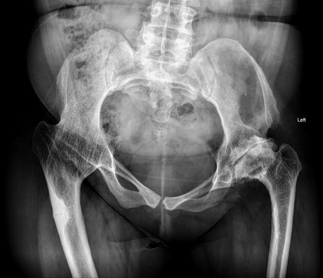
Radiographic features
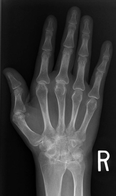
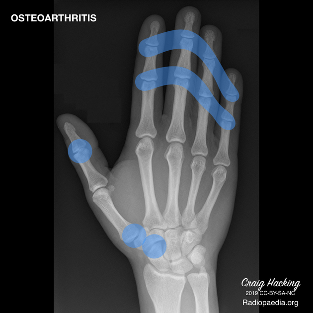
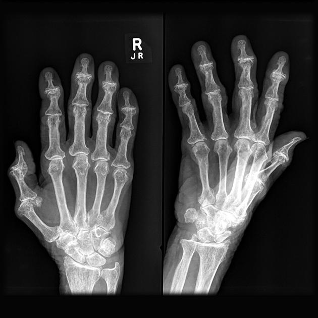
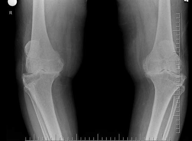
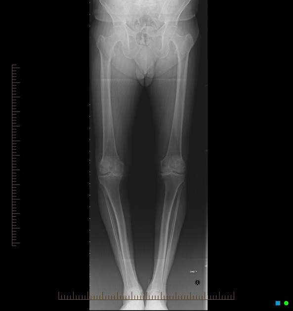
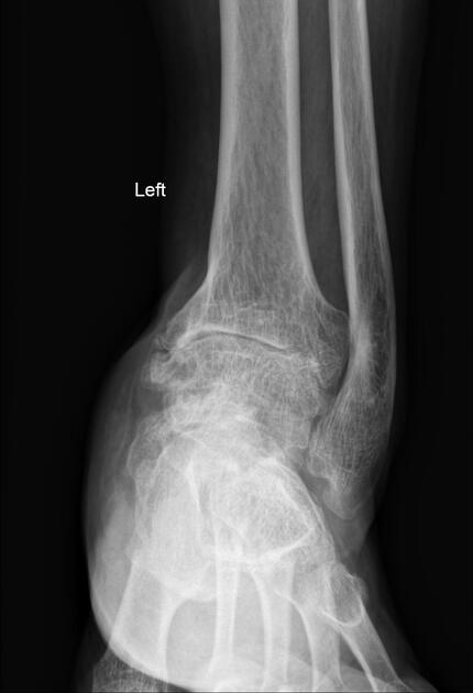
Key radiographic features are joint space narrowing, subchondral sclerosis, and osteophytosis. If all three of these findings are not present, another diagnosis should be considered. With the increasing use of MRI in the assessment of osteoarthritis, other findings have been studied, such as bone marrow lesions and synovitis.
-
joint space narrowing
characteristically asymmetric
least specific: present in many other pathological processes
-
subchondral sclerosis
sclerotic changes occur at joint margins
frequently seen unless severe osteoporosis is present
-
osteophytosis
i.e. development of osteophytes
common degenerative joint disease finding
will also be diminished in the setting of osteoporosis
some osteophytes carry eponymous names: e.g. Heberden nodes, Bouchard nodes
-
joint erosions
-
several joints may exhibit degenerative erosions 1:
-
-
subchondral cysts
also known as geodes
cystic formations that occur around joints in a variety of disorders, including, rheumatoid arthritis, calcium pyrophosphate dihydrate crystal deposition disease (CPPD), and avascular necrosis
-
bone marrow lesions 14,16
visible on MRI as bone marrow oedema-like lesions, often adjacent to areas of cartilage damage - likely representing early osteoarthritis changes
have been shown to correlate with joint pain and progression of cartilage loss
may progress to subchondral cysts
-
a non-specific finding, present also in other diseases, including inflammatory and infectious conditions
present in up to 50% of patients with osteoarthritis 14
according to some authors it may be correlated with pain, disease severity and progression 14,15
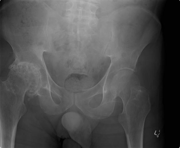
Plain radiograph
Plain radiograph is the most commonly used modality in assessment of osteoarthritis due to its availability and low cost. It can detect bony features of osteoarthritis, such as joint space loss, subchondral cysts and sclerosis, and osteophytes. It is, however, relatively insensitive to early disease changes. Other limitations are a lack of assessment of soft-tissue structures and low intrareader reliability 17.
Scoring systems used to assess the severity of osteoarthritis on radiographs include 17,18:
Osteoarthritis Research Society International (OARSI) atlas
Ultrasound
Ultrasound is not routinely used in osteoarthritis. The assessment of the bony structure and deep joint structures using this modality is impossible. However, it is useful in detecting joint effusion, synovitis, and osteophytes. It can also act as guidance in joint interventions.
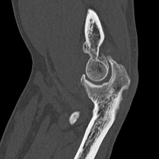
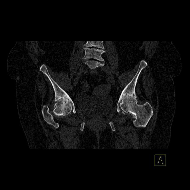
CT
CT has excellent accuracy in assessing bony osteoarthritis changes. It is especially useful in the assessment of the facet joints. In order to reliably assess the articular cartilage, an arthro-CT must be performed.
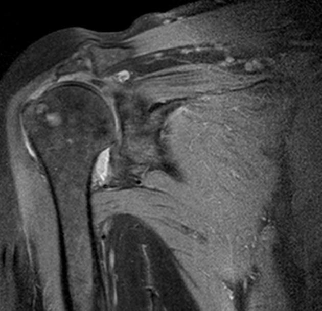
MRI
MRI can very accurately assess both bones and soft-tissue joint structures. It can detect bone marrow changes and cartilage loss, both of which are early osteoarthritis changes and are not visible on radiographs. On conventional MR, articular cartilage is best assessed using fluid-sensitive sequences with fat suppression; with the advent of methods of cartilage quantification and composition assessment - used in research - the sensitivity is further increased. Contrast administration enhances the visualisation of synovitis.
Several scoring systems using MRI assessment of osteoarthritis of the knee have been proposed 14:
Whole-Organ Magnetic Resonance Imaging Score
Knee Osteoarthritis Scoring System
Boston Leeds Osteoarthritis Knee Score
Nuclear medicine
While not routinely used in clinical practice, nuclear medicine studies can provide information about multiple joints in one examination. The changes in the joints with osteoarthritis show increased radiotracer uptake due to reactive bone turnover. The potential disadvantage of a poor anatomical resolution can be solved by using hybrid imaging 14.
Nuclear medicine examination used in the assessment of osteoarthritis are:
scintigraphy with Tc-99m hydroxymethylene diphosphonate (HDP)
PET with 18FDG or 18F
Treatment and prognosis
There is no effective treatment to slow or reverse the changes of osteoarthritis 7. The mainstays of treatment include exercise, walking aids, bracing, and analgesia (including intra-articular steroid injections) 8. Arthroplasty can result in improved function and reduced pain 10.
History and etymology
The term "osteoarthritis" was introduced as a synonym for rheumatoid arthritis by the English physician John K Spender (1829-1916) in 1886, however, it was not until 1907 that the English physician Archibald E Garrod (1857-1936) applied the term to the condition that is now considered to be osteoarthritis 19-21.


 Unable to process the form. Check for errors and try again.