
Paramagnetic rim lesions, less commonly known as iron rim lesions, are neuroimaging biomarkers typically seen in multiple sclerosis, and are included in the McDonald criteria.
Paramagnetic rim lesions are typically seen in and are specific for multiple sclerosis, where they may be seen in approximately 50% of patients with either relapsing or progressive disease 1,3,4. They reflect chronic low-grade (i.e. smoldering) inflammation with iron accumulation in microglia (macrophages) at the lesional edge 1-4. Iron is a paramagnetic substance, thus, these lesions demonstrate a hypointense rim on susceptibility-weighted sequences 3. Presence of paramagnetic rim lesions correlates with increased disability in multiple sclerosis 1,3,4.
On this page:
Radiographic features
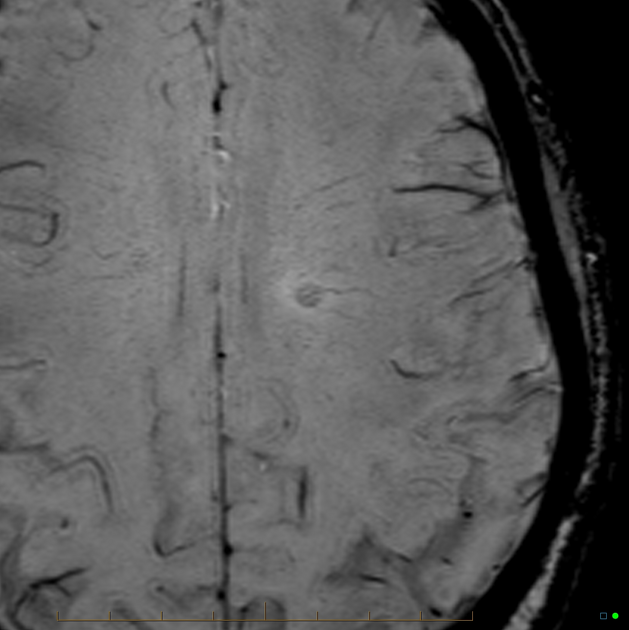
Paramagnetic rim lesions are seen on susceptibility-weighted MRI sequences (e.g. SWI, SWAN, T2*), and co-register with high T2/FLAIR signal lesions 1-4. They derive their name from having a hypointense rim, as seen on susceptibility-weighted MRI, surrounding an internal lesion that is isointense to adjacent, normal, extralesional white matter 1-4. Some descriptions of paramagnetic rim lesions propose that the hypodense rim should involve at least 50% of the lesion’s total rim 2.
These lesions were originally described on 7 T MRI, however, can be readily and accurately appreciated on more conventional and available 1.5 and 3 T strength MRI as well 1,2.
Differential diagnosis
Paramagnetic rim lesions are not pathognomonic of multiple sclerosis as they may very rarely be seen in other central nervous system pathologies 1. For example, they have been described to rarely occur in Susac syndrome, tropical spastic paraparesis, neuromyelitis optica spectrum disorder, and Sjögren syndrome 1. The pathogenesis of the paramagnetic rim is likely different in these pathologies, for example, in Susac syndrome this may be due to hemoglobin extravasation rather than chronic low-grade inflammation 1.


 Unable to process the form. Check for errors and try again.