
The term pelvis (plural: pelvises or pelves) can refer to either the bony pelvis or the pelvic cavity.
On this page:
Bony pelvis
The bony pelvis is formed by the sacrum and coccyx and a pair of hip bones ("ossa coxae"), which are part of the appendicular skeleton. Its primary function is the transmission of forces from the axial skeleton to the lower limbs as well as supporting the pelvic viscera.
Until puberty, each hip bone consists of three separate bones yet to be fused: ilium, ischium and pubis connected by the triradiate cartilage.
The two hip bones are joined anteriorly at the pubic symphysis and posteriorly to the sacrum at the sacroiliac joint. The hip bones incorporate the acetabulum, which articulates with the proximal femur at the hip joint.
Sex differences
Differences between the males and female bony pelvis arise as an adaptation of the female pelvis to childbearing 3:
- infrapubic angle is greater than 90° in females
- pelvic inlet shape
- males: heart-shaped
- females: round or oval
- wider greater sciatic notch in females
- acetabulum faces more anteriorly in females
- sacrum more triangular and shorter in females
- oval or triangular obturator foramen in females
The shape of the female bony pelvis can be described using the following terms 3:
- gynaecoid pelvis (50%): normal female type
- anthropoid pelvis (25%): long AP diameters, short transverse diameters and narrow infrapubic angle
- android pelvis (20%): male type with conical shaped pelvic cavity and heart-shaped pelvic inlet
- platypelloid ("flat female") pelvis (5%): short AP diameters, long transverse diameters and wide infrapubic angle
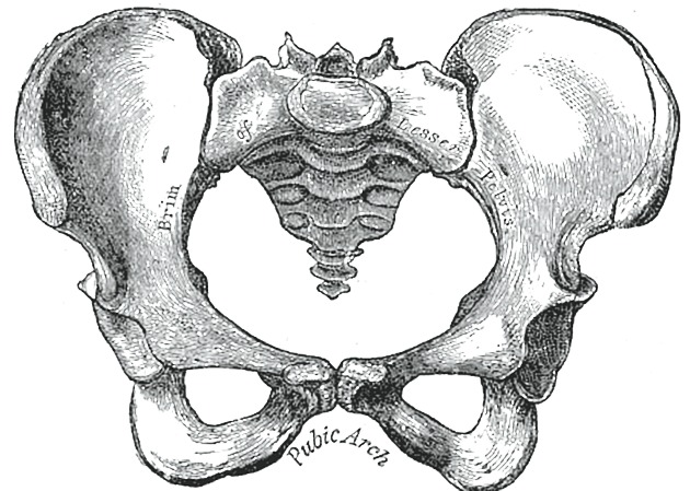
Pelvic apertures
The pelvic brim defines the pelvic inlet and the following structures contribute to it 2:
- anteriorly: pubic crest, pecten pubis
- laterally: arcuate line (of the ilium)
- posteriorly: sacral ala
The pelvic outlet is formed by the following structures 2:
- anteriorly: pubic arch, inferior margin of the pubic symphysis, pubic rami, ischial rami
- laterally: ischial tuberosities
- posterolaterally: sacrotuberous ligaments
- posteriorly: sacrum and coccyx
Pelvic cavity
The pelvic cavity is inferior part of the abdominopelvic cavity and is in direct connection with the abdominal cavity.
The pelvic cavity is bounded by the bony pelvis and the pelvic musculature and primarily contains reproductive organs and the rectum. It is divided into:
- lesser pelvis (or true or minor pelvis) is inferior to the pelvic brim and lies between the pelvic inlet and the pelvic outlet
- greater pelvis (or false or major pelvis) is the expanded portion of the cavity situated superior and anterior to the pelvic brim
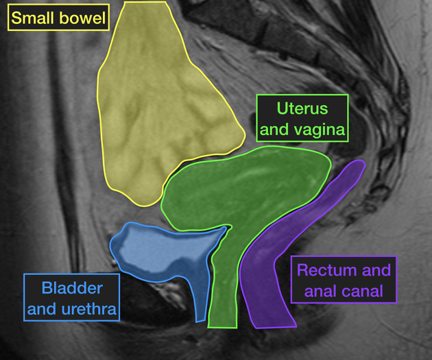
Pelvic viscera
The visceral contents of the pelvis is different between the sexes due to the difference in gonadal descent and gonadal tube formation 2.
- common: rectum, urinary bladder, ureters
- male: prostate, male urethra, seminal vesicles, ejaculatory ducts, ductus deferens
- female: ovaries, uterine tubes, uterus, vagina, female urethra
Radiographic features
Plain radiograph
Useful for imaging the bony pelvis and can be used in contrast studies, for example, urethrograms or intravenous pyelograms (IVP).
Ultrasound
First-line imaging modality for assessing both the male and female pelvic viscera. Doppler ultrasound can also be used to assess the pelvic vasculature 3.
CT
Due to radiation exposure to the gonads, CT is not the first line imaging test for evaluating the pelvic viscera except in the setting of trauma 3.
MRI
MRI provides superior soft tissue contrast resolution for imaging the anatomy (best seen in T1-weighted) and pathology (best seen on T2-weighted) of the pelvis 3.
Angiography
Invasive angiography is the gold standard modality for assessing pelvic vasculature 3.


 Unable to process the form. Check for errors and try again.