
Penile implants are surgically placed devices to assist with erectile dysfunction. The device is comprised of either two malleable rods or inflatable components inserted in the penile shaft with or without a pump/reservoir located in the scrotum or pelvis.
On this page:
Types
-
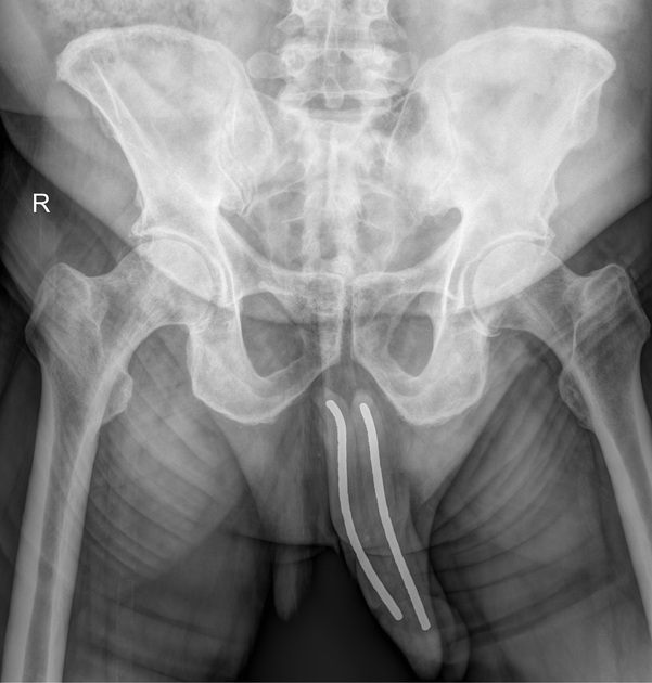
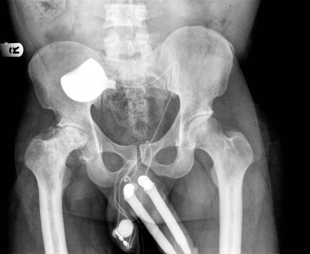
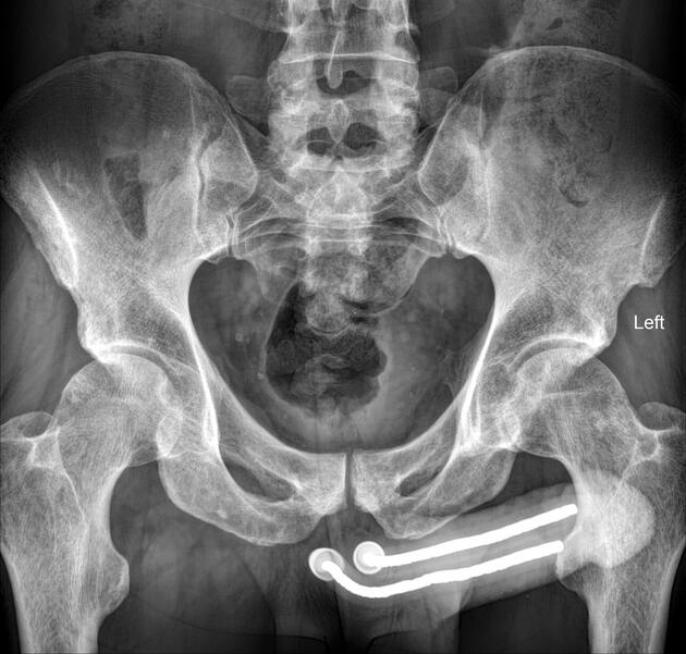
semirigid or malleable penile prostheses (MPP)
paired malleable rods that are inserted surgically into each corpora cavernosa 2
usually made of stainless steel or a braided silver core coated in silicone 2
more prone to lateral perforation and distal erosion 2
-
hydraulic or inflatable penile prostheses (IPP)
-
two types
two-piece: consists of two cylinders within the corpora cavernosa and a resipump (combined pump and reservoir) placed in the scrotum 2
three-piece: consists of two cylinders within the corpora cavernosa, a pump in the scrotum with a reservoir placed adjacent to the bladder 2
primarily non-metallic (unlike MPPs) excluding a rear tip extender component at the base that guarantees stability 3
more popular as offers the best flaccidity with rigidity nearing that of a natural erection, placement however is surgically complex 2
-
Radiographic features
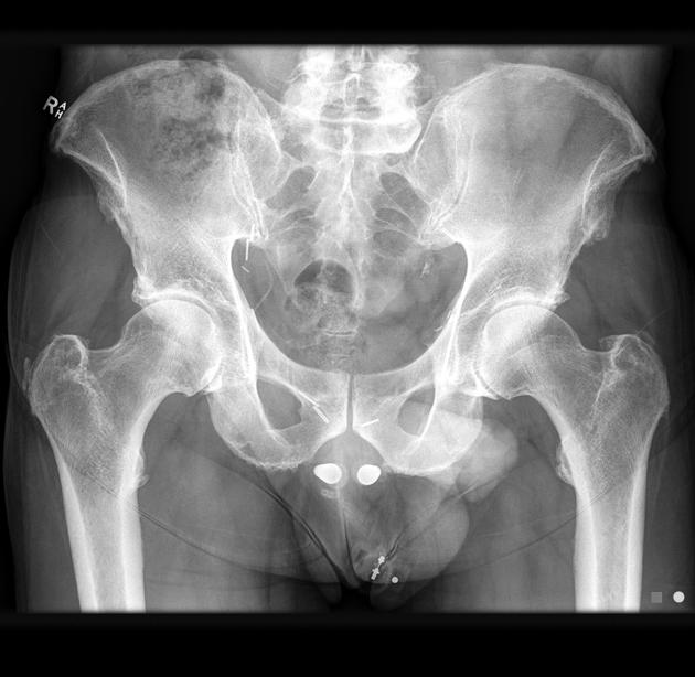
Plain radiograph
Plain films are only capable of identifying complications of a mechanical nature such as fractures and torsions of MPPs due to their metallic core.
As IPPs have become more mainstream and their only metallic component is the rear tip extender at the base, plain film has become an outdated imaging modality 3. Historically IPPs were filled with radiopaque material allowing visualization on plain film but this is no longer the case as saline solution is used 2.
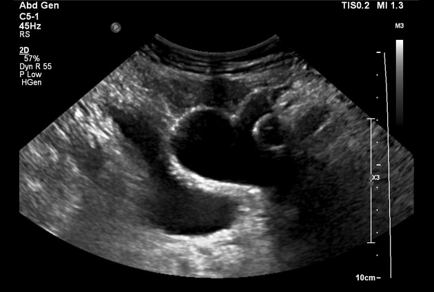
Ultrasound
Complications such as reservoir migration, blockage and leakage of IPP implants can be diagnosed on ultrasound imaging 3.
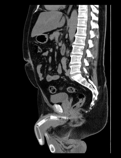
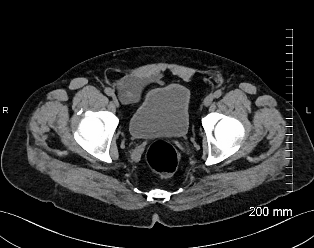
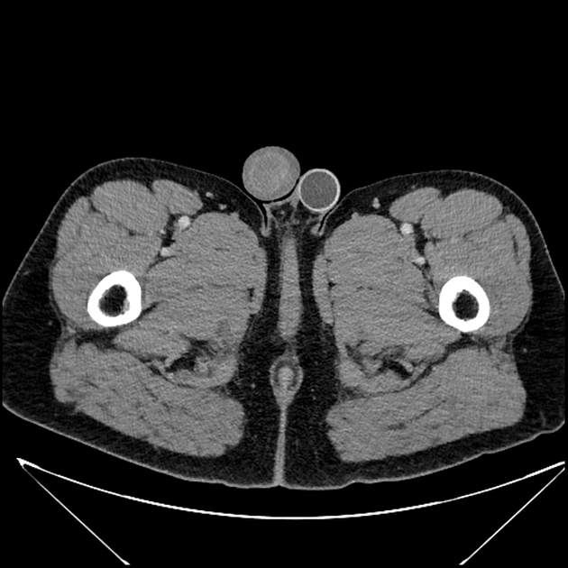
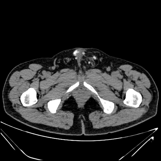
CT
While both MPPs and IPPs are visible on CT, internal architecture is not easily visible and position within the corpora cavernosa cannot be confidently assessed 3. Infections, fractures and hematomas can be assessed emergently however due more widespread access to CT imaging 3.
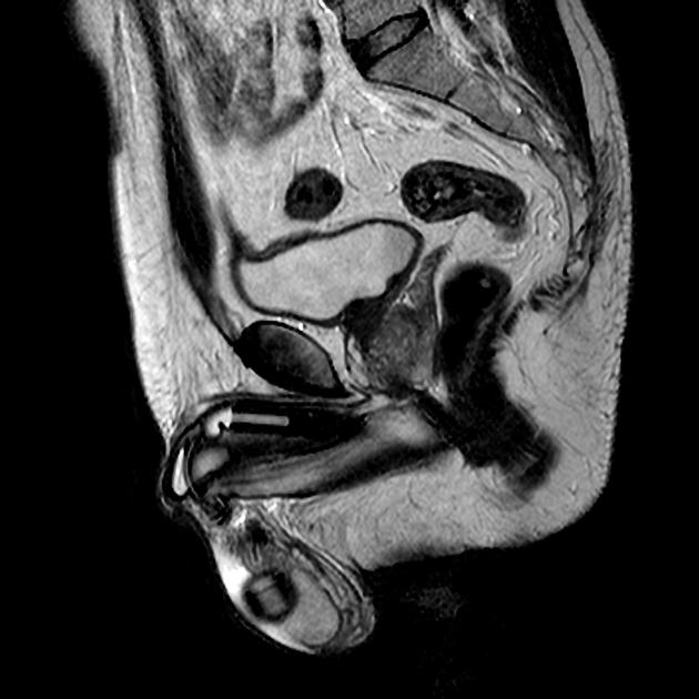
MRI
MRI is the modality of choice for investigation of patients with suspected penile prosthesis complications 3. When positioning the patient, they should be placed in the supine position with a towel placed between the legs to ensure elevation of the penis and scrotum 3. The penis itself can also be taped to the anterior abdominal wall.
MPPs appear hypointense on T1 and T2 imaging due to low signal intensity of the metallic core and it's silicone cover 3. Some models may have articulated segments that may be identified by heterogenous signal intensity 3. This is significant as fractures can occur along the shaft 3.
IPPs appear T2 hyperintense due to their volume of saline solution. The silicone base that covers the cylinders is visible as a T2 hypointense layer outlining their shape 3.
Complications
leak around the the tubes or the reservoir, with subsequent infection
migration of the fluid balloon reservoir along its track and may get lodged anywhere between the muscle and the skin - similar to a buried bumper syndrome
History and etymology
They were thought to have been first introduced by Beheri et al in the 1960s 2.


 Unable to process the form. Check for errors and try again.