
Percutaneous endoscopic gastrostomy (PEG) is a procedure where a flexible feeding tube (commonly known as a PEG tube) is inserted through the abdominal wall and into the stomach via endoscopy. Alternatively a tube can be placed under radiological guidance, known as a radiologically inserted gastrostomy (RIG) or percutaneous radiological gastrostomy (PRG).
A PEG tube permits nutrition, fluids and/or medications to be placed directly into the stomach, bypassing the mouth and esophagus. This is typically performed when a patient is either unable to, or it is unsafe for them to, consume nutrition orally (e.g. in patients with swallowing issues or head and neck malignancies).
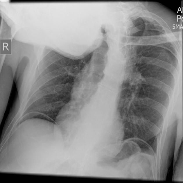
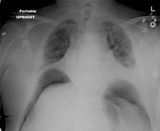
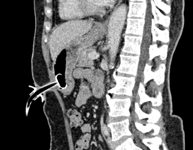
On occasion it is necessary to image for complications of PEG tubes - either immediately after insertion or when long standing. The typical clinical problem relates to concerns over displacement, including buried bumper syndrome.
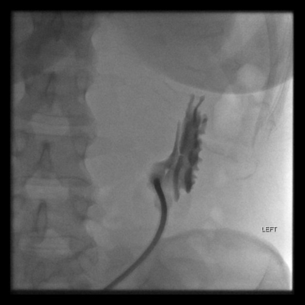
Occasionally contrast will be injected via the PEG to determine its position, a so-called PEGogram.
History and etymology
PEG was originally co-developed by two Americans, pediatric surgeon, Michael Gauderer, and endoscopic surgeon Jeffrey L Ponsky, both of whom worked at the Rainbow Babies and Children's Hospital in Cleveland, Ohio. The first PEG was performed in 1979 3.


 Unable to process the form. Check for errors and try again.