
This is a basic article for medical students and other non-radiologists
Pneumonia refers to infection within the lung and results in infective fluid and pus filling the alveolar spaces. This initially results in patchy airspace opacification and then more confluent consolidation.
On this page:
Reference article
This is a summary article; read more in our article on pneumonia.
Summary
- anatomy
-
epidemiology
- a wide group of affected individuals
-
presentation
- productive cough
- shortness of breath
- chest pain
-
pathophysiology
- the infection causes exudate to accumulate in the lung
- this exudate fills the alveolar spaces
- initially, this is incomplete
- as the infection worsens, all the air is displaced, and the alveoli are filled with infection
-
investigation
- blood workup
- raised white-blood-cell count and inflammatory markers
- chest x-ray
- first line investigation
- confirm infection
- assess severity and complications
- CT chest
- further assessment of complications
- assessment of causes in recurrent (non-resolving) infection
- blood workup
-
treatment
- in most cases, antibiotic therapy is all that is required
- this may be oral or intravenous, depending on the severity
- the Pneumonia Severity Index (PSI) or the more simplified CURB-65 score may be used for risk stratification
- in most cases, antibiotic therapy is all that is required
Radiographic features
Chest radiograph
- airspace opacification
- filling of the alveoli with infectious material and pus
- initially patchy
- becomes confluent as infection develops
- air bronchograms
- air-filled bronchi running through pus-filled alveoli
- complications
- pleural collection
- cavitation
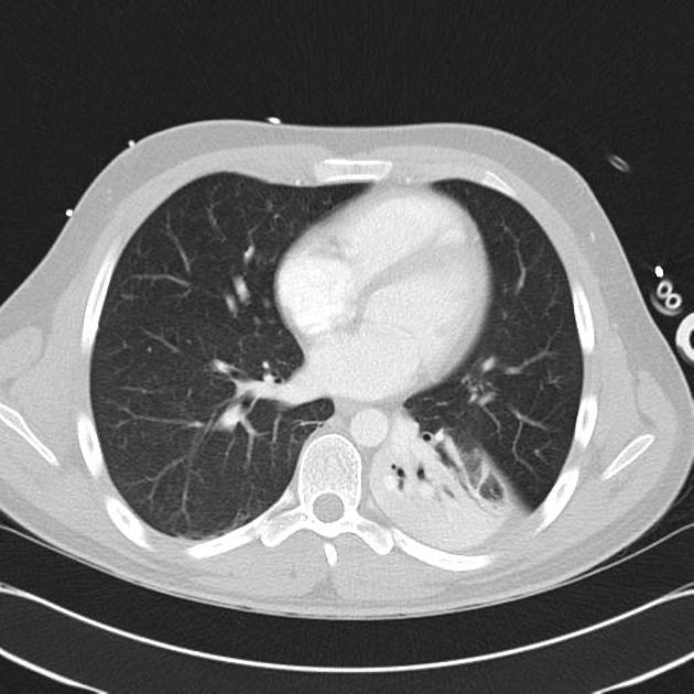
CT chest
- airspace opacification
- looks the same as on a chest x-ray
- degree of consolidation assessed more accurately
- complications can be seen earlier than a chest x-ray
- lung necrosis
- cavitation
- empyema


 Unable to process the form. Check for errors and try again.