
The popliteal artery is the direct continuation of the superficial femoral artery, at the point where it exits the adductor canal at the adductor hiatus, and passes into the popliteal fossa as the vessel courses posteriorly behind the knee.
On this page:
Summary
origin: continuation of the superficial femoral artery as it exits the adductor canal
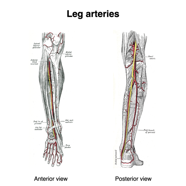
main branch: anterior tibial artery
termination: continues as the tibioperoneal trunk in the lower aspect of the popliteal fossa
Gross anatomy
Origin
As a continuation of the femoral (superficial femoral) artery as it passes into the popliteal fossa through the adductor canal at the junction of the middle and lower thirds of the thigh.
Course
Descends as the deepest structure on the floor of the popliteal fossa. Courses just superficial to the popliteus muscle.
For surgical or angiographic purposes, the popliteal artery can be divided into three segments 4:
P1: from adductor hiatus to the top of the patella
P2: from top of the patella to center of the knee joint
P3: from center of the knee joint to anterior tibial artery origin
Termination
At the lower border of the popliteus, divides into its terminal branches - anterior tibial artery and tibioperoneal trunk.
Branches
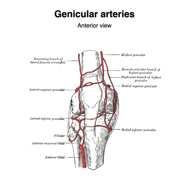
superior medial and lateral genicular arteries - course around the femoral condyles as part of the genicular anastomosis
inferior medial and lateral genicular arteries - course around the tibial condyles as part of the genicular anastomosis
middle genicular artery - pierces oblique popliteal ligament
muscular branches - supply hamstrings, adductor magnus, and triceps surae
cutaneous branches - supply skin over knee joint
Relations
popliteal artery starts medial to tibial nerve and ends lateral to it; the popliteal vein always between the two
deep/anterior: popliteal surface of the femur (separated by fat), knee joint capsule, and popliteus
superficial/posterior: popliteal vein, tibial nerve (most superficial)
lateral: (above) biceps femoris and lateral condyle of the femur, (below) plantaris and lateral head of the gastrocnemius
medial:(above) semimembranosus and medial condyle of the femur, (below) medial head of the gastrocnemius
Variant anatomy
Variants in popliteal branching pattern 1,2:
popliteal artery arising off a persistent sciatic artery
~90% of the population displays the typical branching pattern of bifurcation into an anterior tibial artery and tibioperoneal trunk
-
~10% have variant branching pattern:
hypoplastic infrapopliteal vessels ~4% (range 1.0-6.8%)
trifurcation (no tibioperoneal trunk) ~2% (range 1.5-3.2%)
high bifurcation (above popliteus)
high origin of anterior tibial artery ~2.5% (range 0.5-4.5%)
high origin of posterior tibial artery ~1%
peroneal artery arising off the anterior tibial artery


 Unable to process the form. Check for errors and try again.