
When issuing an MRI or CT report on a patient with an aortic aneurysm, whether it be thoracic or abdominal, several features should be mentioned to aid the referring clinician in managing the patient.
Radiology report
Reporting tips for aortic aneurysms include 1,2:
-
size and shape
sac dimensions (outer surface to outer surface)
luminal diameter if mural thrombus is present
length 3
fusiform or saccular
size of vessel proximal and distal to aneurysm
-
characteristics of wall
mural calcification
presence of mural thrombus
-
location and relationship to involved branches/structure
-
involvement of the origins of the renal arteries
presence of multiple renal arteries and where they arise
great vessels from the arch
-
-
characterization of possible etiology
true or false
possibility of mycotic etiology
-
complications
leak
proximity to bowel
-
other relevant vessels
-
the size and dominance of vertebral arteries should be included if the aneurysm is close to the left subclavian artery
presence of carotid disease is important, as significant stenosis may predispose the patient to strokes during any period of reduced flow / hypotension
-
abdominal aortic aneurysm (AAA)
renal veins: preoperative knowledge of the presence of variant anatomy is essential e.g. retroaortic or circumaortic left renal veins
access vessels for endovascular repair (EVAR) which usually requires bilateral common femoral artery (CFA) punctures and vessel diameters of 6 mm or greater
-
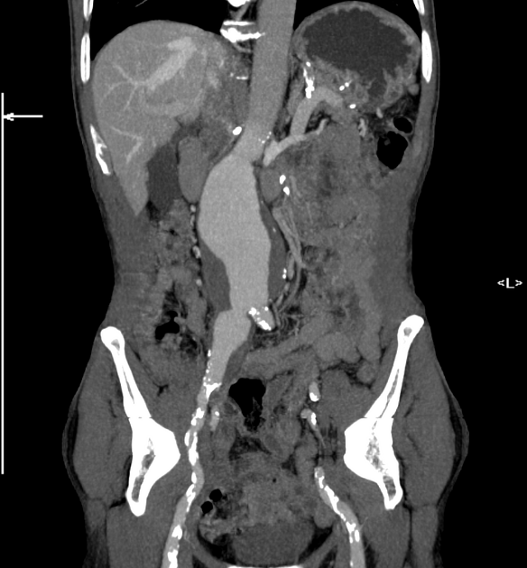
Example
The infrarenal aorta demonstrates a fusiform aneurysm which measures 6 x 7 cm (AP x LR) and extends over a distance of 7.5 cm. It begins 2.3 cm distal to the renal arteries (aorta at this level measures 2.1 cm) and terminates at the aortic bifurcation without extension into the iliac arteries (right CIA = 1.2 cm, left CIA = 1.1 cm).
Smooth eccentric mural thrombus is present within the aneurysm, thickest on the right where it measures 1.5 cm. The lumen has a diameter of 4.5 x 6 cm (AP x LR). There is no surrounding stranding or extravasation of contrast to suggest impending rupture.
The aorta and other visualized vessels demonstrate widespread atherosclerosis, suggesting that this is the underlying etiology of the AAA. Moderate (<50%) stenosis is seen at the origin of the right renal artery and celiac trunk.
The common femoral arteries bilaterally demonstrate calcific atherosclerosis but no stenosis. High-grade stenosis is however present in the right external iliac artery, potentially posing problems for endovascular access on this side.


 Unable to process the form. Check for errors and try again.