
Getting a film with right upper lobe collapse in the exam is one of the many exam set-pieces that can be prepared for.
On this page:
Description
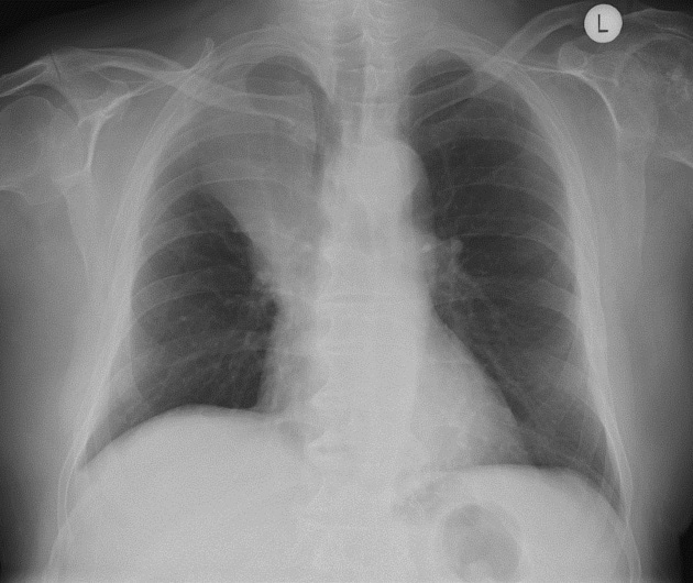
There is increased opacification in the right upper zone with associated volume loss - rib spacing is reduced, midline structures displaced to the right and the right hilum and right hemidiaphragm are elevated.
There is increased density at the right hilum consistent with a mass causing upper lobe collapse.
Review of the remainder of the lung fields and bones is normal. The costophrenic angle is crisp and there is no suggestion of pleural fluid on either side.
Further investigation with a staging CT scan of the chest and upper abdomen is suggested to confirm the presence of a primary obstructing bronchial carcinoma and stage the primary tumor, nodal disease and distant metastases.
Notes
know the TNM staging of lung cancer


 Unable to process the form. Check for errors and try again.