
Splenic artery aneurysms are the most common visceral arterial aneurysm formation as well as the third most common abdominal aneurysm (after the aorta and iliac vessels). Aneurysms are usually saccular in configuration and they can either be in the form of a true aneurysm (much more common) or as a pseudoaneurysm.
This article focuses on the true splenic artery aneurysm, please refer to splenic artery pseudoaneurysms for a specific discussion on this entity.
On this page:
Epidemiology
The true prevalence of splenic artery aneurysms is unknown. Estimates vary widely from 0.2% to 10.4%, but generally, it is the third most common site of intra-abdominal aneurysms after abdominal aorta and iliac arteries 1,6. Incidentally discovered splenic artery aneurysms are being diagnosed more frequently with wider use of cross-sectional imaging modalities 7.
Splenic artery aneurysms are about 4x more common in females, yet the risk of its rupture is about 3x more common in males 13.
Associations
Clinical presentation
Most splenic artery aneurysms are silent and are discovered in asymptomatic patients 2. More than 95% of patients with non-ruptured splenic artery aneurysms were asymptomatic 13. The risk of rupture increases with liver transplantation, portal hypertension, and pregnancy. A ruptured splenic artery aneurysm usually presents with sudden onset left upper quadrant abdominal pain followed by hemodynamic instability, and gastrointestinal bleeding 14.
The so-called “double rupture” phenomenon occurs when initial spontaneous stabilization followed by subsequent sudden circulatory collapse is experienced. This is caused by initial bleeding, collecting into the lesser sac then followed by flooding into the peritoneal cavity 15.
Pathology
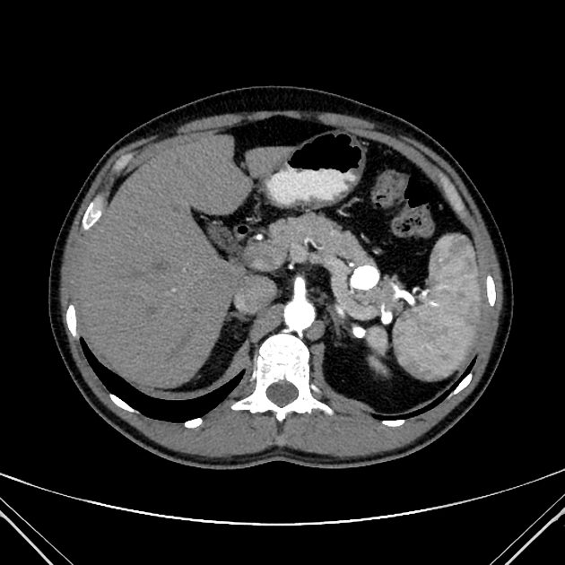
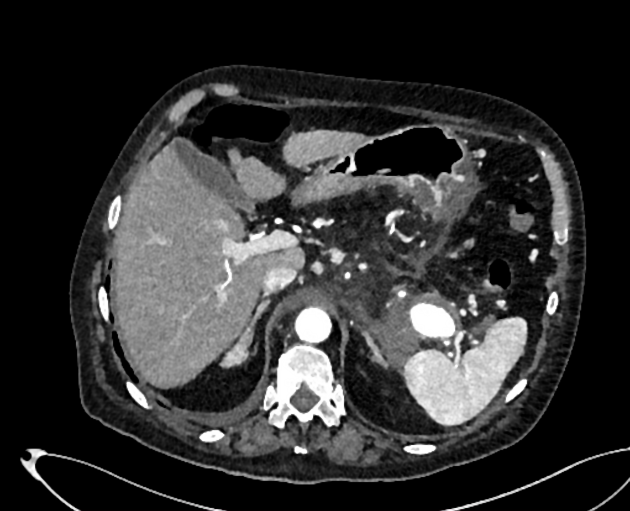
The size of splenic artery aneurysms can range from 2 to 9 cm, but usually, they are smaller than 3 cm. Those may be single or multiple and are most commonly involving the distal portion of the artery. Peripheral calcification is common, and mural thrombus may be present 12.
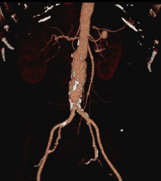
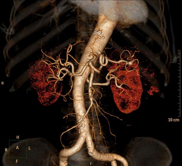
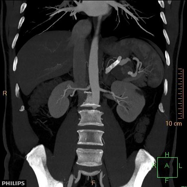
Radiographic features
Several imaging modalities can be used to diagnose splenic artery aneurysms, with CTA the recommended modality 18.
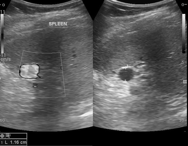
Ultrasound
Ultrasound with Doppler is a non-ionizing modality and relatively cheap and available yet it is operator dependent, has limited spatial resolution and may be difficult in cases of obesity, bowel shadowing and atherosclerosis 18. It has poor sensitivity for detecting aneurysms <3 cm 18.
CT
CT angiography is a quick and efficient examination, has a higher resolution than MR and its MPR capabilities greatly help in diagnosis, yet it has its contraindications (renal failure, allergy to contrast).
MRI
MR angiography is of higher spatial resolution than ultrasound but it is relatively expensive and contraindicated in some patients (claustrophobia, aneurysm clips, cardiac pacemakers).
Angiography (DSA)
Direct catheter angiography is the gold standard imaging test, has the highest resolution and concomitant management can be applied, yet it still carries the complications of arterial puncture 8-11.
Treatment and prognosis
The overall risk of rupture is thought to be ~6%. However, in an event of rupture, there is a relatively high mortality rate of ~ 30% (range 25-36%) 4,18. Pregnancy is a risk factor for rupture, particularly in the third trimester, with very high maternal (80%) and fetal (90%) mortality rates 18.
The Society for Vascular Surgery 2020 guidelines recommend 18:
-
surgical management of
ruptured splenic artery aneurysms
splenic artery pseudoaneurysms of any size
splenic artery aneurysms of any size in patients who have childbearing potential
non-ruptured splenic artery aneurysms ≥3 cm with a demonstrable increase in size or symptomatic
-
observation of splenic aneurysms <3 cm in asymptomatic patients or those unfit for surgery
annual CT or ultrasound surveillance
Surgical management may consist of open or laparoscopic repair, splenic artery ligation +/- splenectomy or endovascular treatment depending on the clinical scenario 18. Periodic CTA surveillance post endovascular treatment is recommended to assess for recurrence 18.
Differential diagnosis
calcified left adrenal hematoma


 Unable to process the form. Check for errors and try again.