
Subependymal hamartomas are seen in patients with tuberous sclerosis. They are located along the ventricles and are mostly asymptomatic. As with other hamartomas, they grow at the same rate as the surrounding tissues.
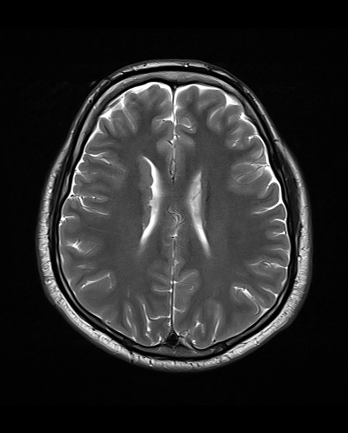
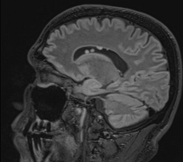
On imaging, they appear as small intraventricular masses, smaller than 1 cm, and demonstrate variable signal on MRI with contrast enhancement, and may calcify.
On this page:
Epidemiology
Subependymal hamartomas are a well-known manifestation of tuberous sclerosis, affecting 80% of patients with the condition 1. They are visible within the first six months of age 2.
Clinical presentation
Subependymal hamartomas are often asymptomatic. When symptoms occur, they are usually a result of obstructive hydrocephalus from the mass effect to the ventricular system.
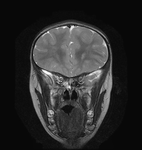
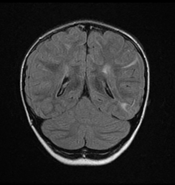
Radiographic features
Subependymal hamartomas are small irregular nodules, measuring <1 cm, with their long axis perpendicular to the ventricular surface. They grow in proportion to the surrounding tissues and may calcify with increasing age.
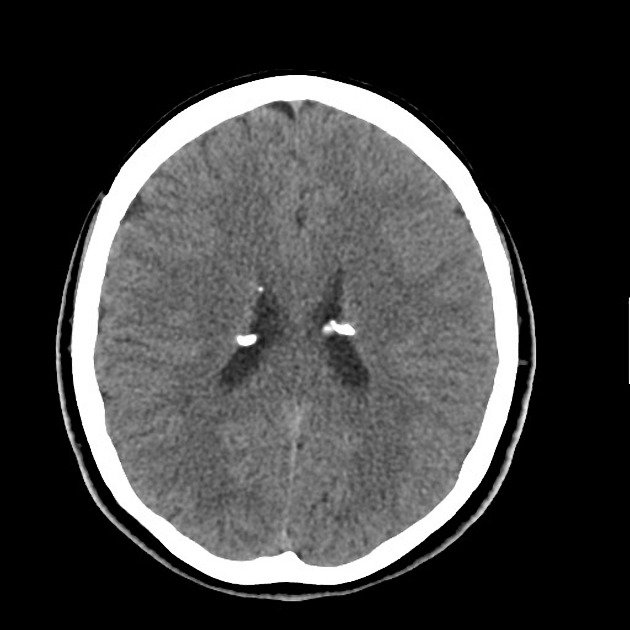
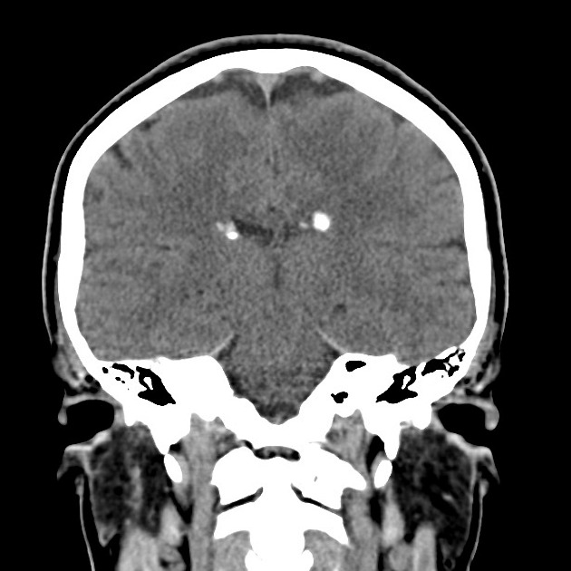
CT
small (less than 1 cm) irregular intraventricular mass
calcification is common (88%)
accompanying hydrocephalus may be present
variable contrast enhancement
MRI
T1: variable signal, frequently hyperintense to grey matter
T2: variable signal, frequently iso to hyperintense to grey matter
T1 C+ (Gd): also shows variable enhancement
Marked hypointense areas are in keeping with calcification
Treatment and prognosis
Subependymal hamartomas are mostly asymptomatic. However, they may progress to subependymal giant cell astrocytoma which may lead to obstructive hydrocephalus, causing morbidity or mortality. Therefore, surveillance is offered to patients with tuberous sclerosis.
Should these tumors become symptomatic or large, surgical treatment is required.
Differential diagnosis
-
subependymal giant cell astrocytoma
larger than 1 cm
increases in size in serial imaging 3,4
-
subependymal grey matter heterotopia
smooth and ovoid
long axis parallel to the ventricular surface
follows grey matter signal
-
ring-shaped lateral ventricular nodules 5
smaller than 1 cm
ring-shaped with a core signal that follows CSF signal intensity
does not increase in size on follow-up


 Unable to process the form. Check for errors and try again.