
Swyer-James syndrome, also known as Swyer-James-MacLeod syndrome and Bret syndrome, is a rare lung condition that manifests as unilateral hemithorax lucency as a result of postinfectious obliterative bronchiolitis.
On this page:
Epidemiology
The condition typically follows a viral respiratory infection such as adenoviruses or Mycoplasma pneumoniae infection in infancy or early childhood 2-6. Non-infectious cause includes ingestion of hydrocarbon. It has an estimated prevalence of 0.01% 9.
Radiographic features
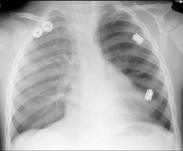
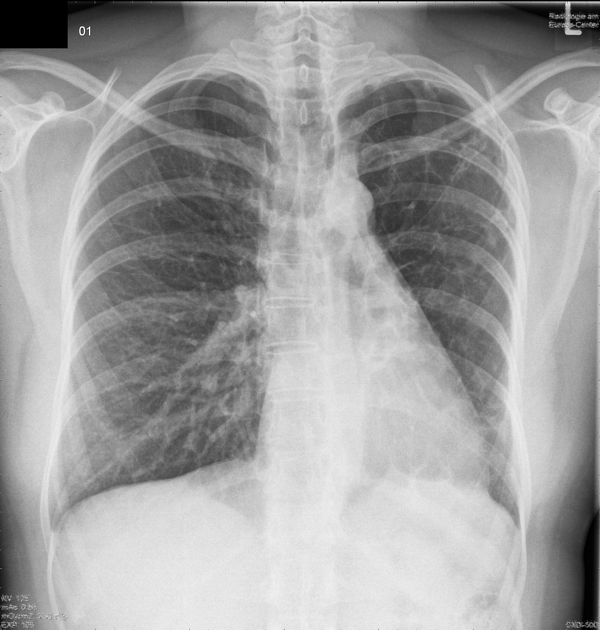
Plain radiograph
It is generally characterized on radiographs by a unilateral small lung with hyperlucency and air trapping on expiration 2.
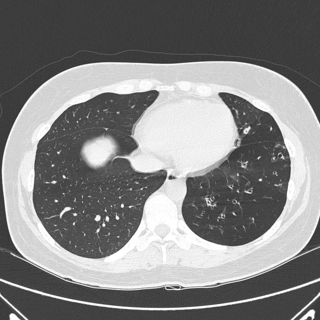
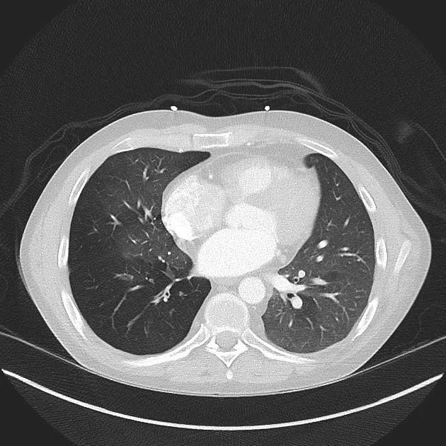
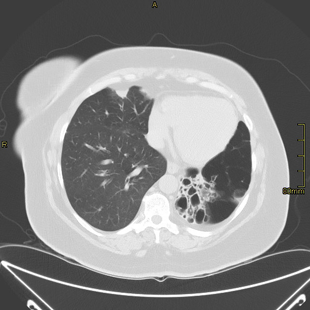
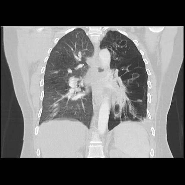
CT
CT shows the affected lung as being hyperlucent with diminished vascularity. The disease can be unilateral or bilateral. The entire lung can be affected, however, there can also be lobar, segmental, and subsegmental involvement in a patchy distribution 2. The size of the majority of the affected lobes is smaller, although occasionally they can be of normal size 3. There is usually no anteroposterior gradient attenuation 4. Bronchiectasis may be present, although this is not a universal finding 5.
MRI
MR angiography (MRA) may show a small pulmonary artery with diminished vascularity in the periphery 8.
Nuclear medicine
Quantitative ventilation-perfusion (V/Q) lung scan shows a photopenic area in the affected aspect.
Treatment and prognosis
Treatment is conservative and preventative, focused primarily on controlling pulmonary infections. Inhaled corticosteroids may have a limited role in treatment 9.
History and etymology
The condition was first described in a case report of a six-year old boy that had been seen in 1949 in Warwick in the UK. The paper was published in 1953 by Paul Robert Swyer (1921-2019 12) a pediatrician, who by that time had emigrated to Canada, and a British radiologist, George C W James 13.
In 1954, an English pulmonologist, William Mathieson MacLeod (1917-1977 11) published a review of nine cases, and cited Swyer and James's paper 14.
It has also been referred to as MacLeod syndrome, but this is not advised given the presence of a rare genetic malformation bearing a similar name: McLeod syndrome.
It was also described by Janus Bret in France in 1956, hence reference to the same condition as Janus syndrome and Bret syndrome 10.
Differential diagnosis
Possible imaging differential considerations include:
congenital lobar overinflation, though the lucent lung, in this case, is usually larger
unilateral absence/proximal interruption of the main pulmonary artery
hypogenetic lung syndrome (scimitar syndrome)
One should also consider other causes of a unilateral hypertranslucent hemithorax.


 Unable to process the form. Check for errors and try again.