Citation, DOI, disclosures and article data
Citation:
Weerakkody Y, Baba Y, Bell D, et al. Thickening of bronchovascular bundles. Reference article, Radiopaedia.org (Accessed on 30 Mar 2025) https://doi.org/10.53347/rID-26071
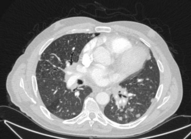
Thickening of bronchovascular bundles is a chest CT imaging feature that can be observed in a number of entities. It has some overlap with the terms peribronchovascular interstitial thickening and peribronchovascular thickening.
Pathology
Causes
Conditions that can result in bronchovascular bundle thickening include:
See also
-
1. Koyama T, Ueda H, Togashi K, Umeoka S, Kataoka M, Nagai S. Radiologic Manifestations of Sarcoidosis in Various Organs. Radiographics. 2004;24(1):87-104. doi:10.1148/rg.241035076 - Pubmed
-
2. Gerke A, van Beek E, Hunninghake G. Smoking Inhibits the Frequency of Bronchovascular Bundle Thickening in Sarcoidosis. Acad Radiol. 2011;18(7):885-91. doi:10.1016/j.acra.2011.02.015 - Pubmed
-
3. Reittner P, Müller N, Heyneman L et al. Mycoplasma Pneumoniae Pneumonia: Radiographic and High-Resolution CT Features in 28 Patients. AJR Am J Roentgenol. 2000;174(1):37-41. doi:10.2214/ajr.174.1.1740037 - Pubmed
-
4. Daimon T, Johkoh T, Sumikawa H et al. Acute Eosinophilic Pneumonia: Thin-Section CT Findings in 29 Patients. Eur J Radiol. 2008;65(3):462-7. doi:10.1016/j.ejrad.2007.04.012 - Pubmed
-
5. Collins C & Quismorio F. Pulmonary Involvement in Microscopic Polyangiitis. Curr Opin Pulm Med. 2005;11(5):447-51. doi:10.1097/01.mcp.0000170520.63874.fb - Pubmed
-
6. Wolfgang Dähnert. Radiology Review Manual. (2011) ISBN: 9781609139438 - Google Books
-
7. Johkoh T, Ikezoe J, Tomiyama N et al. CT Findings in Lymphangitic Carcinomatosis of the Lung: Correlation with Histologic Findings and Pulmonary Function Tests. AJR Am J Roentgenol. 1992;158(6):1217-22. doi:10.2214/ajr.158.6.1590110 - Pubmed
-
8. Ko J, Girvin F, Moore W, Naidich D. Approach to Peribronchovascular Disease on CT. Semin Ultrasound CT MR. 2019;40(3):187-99. doi:10.1053/j.sult.2018.12.002 - Pubmed
Promoted articles (advertising)


 Unable to process the form. Check for errors and try again.