
Thoracic (or pulmonary) histoplasmosis refers to pulmonary manifestations from infection with the fungus Histoplasma capsulatum which is an organism endemic to the Central American state of El Salvador but can be found widely in other parts of both North and South America. It can have variable clinical and radiographic presentations depending on the state of infection and other host co-morbidities.
On this page:
Epidemiology
It is considered the most common systemic fungal infection in North America 3. Most of these are thought to be sporadic in nature, although outbreaks have been reported.
Some populations studies have shown that over 80% of young adults from endemic regions of North America have been previously infected by Histoplasma capsulatum 4.
Histoplasmosis is also present to a lesser extent in Subsaharan Africa, the central Mediterranean, Southeast Asia and Eastern Australia 4.
Clinical presentation
Features interrogated in the history should include travel to endemic areas as well as exposures to significant populations of bats and birds.
-
acute pulmonary histoplasmosis
90% of cases can have no symptoms
symptoms manifest for up to two weeks after exposure
fever, malaise, headache, myalgia, polyarthralgia and abdominal pain
dyspnea can manifest if there is diffuse involvement
cough and hemoptysis can be prominent features if there is a degree of airway compression
rarely dysphagia occurs with esophageal compression
-
subacute pulmonary histoplasmosis
small calcified nodules
hilar or mediastinal lymphadenopathy
-
disseminated progressive histoplasmosis
more likely in patients with deficient cell-mediated immunity including those with AIDS (CD4 <150 cell/μL) or on corticosteroids
can often be fatal
Radiographic features
Most patients with thoracic histoplasmosis are thought to have normal chest radiographs. Where there are findings, these can be non-specific to a range of infectious or inflammatory disorders and histoplasmosis is considered in the differential if the patient is known to have traveled in endemic regions.
The particular imaging features are related to the timeline of the presentation and the patient's ability to mount an immune response.
-
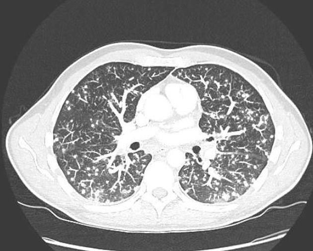
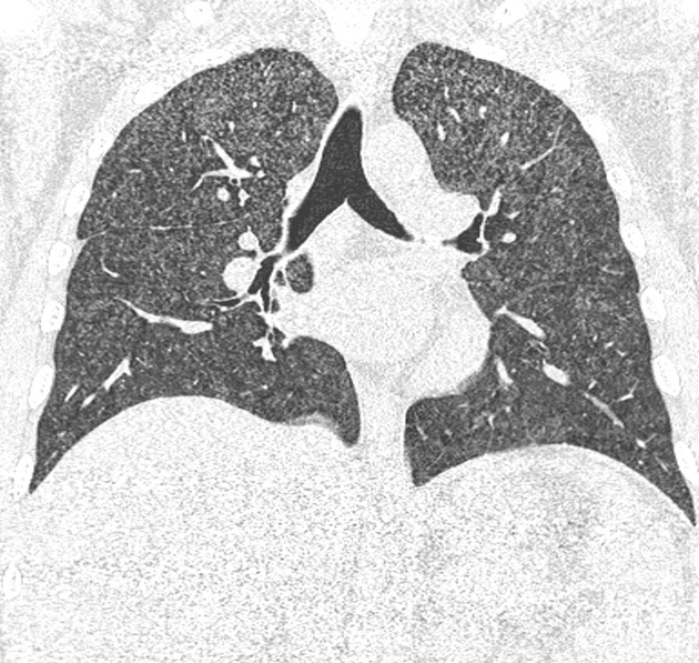
well-defined nodules which have a central calcification giving them a "target lesion" which is pathognomonic
Acute histoplasmosis
airspace shadowing with consolidation involving multiple lung segments or lobes similar to bacterial pneumonia
pleural effusions are possible but uncommon
healing process with formation of histoplasmomas, which are well-defined nodules with central calcifications


 Unable to process the form. Check for errors and try again.