
Presentation
Work up for abdominal pain, dyspepsia, and vomiting from three months ago. Recent breath shortness.
Patient Data


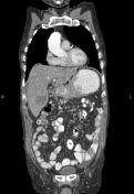
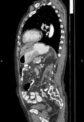
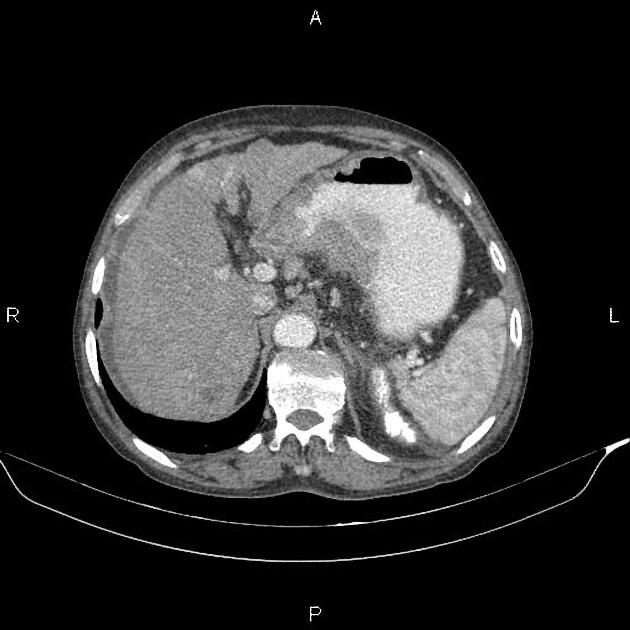
There is increased wall thickness due to tumoral infiltration at the gastric lesser curvature, accompanied by multiple perigastric and porta hepatis enlarged lymph nodes with SAD less than 18 mm.
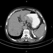
Additionally, multiple hetero enhancing masses are observed throughout the liver with a maximum diameter of 60 mm consistent with metastases.
A small amount of ascites is also evident.
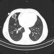
Several nodules, mostly subpleural, are seen in both lungs less than 12 mm. A few calcified granulomas are also evident in lung fields.
Filling defects are seen in the main pulmonary artery branches bilaterally, inferring thromboemboli.
A few nonenhanced simple cortical cysts are seen in both kidneys.
The prostate gland is enlarged, and its central portion protrudes cephalad into the urinary bladder base.
Degenerative changes such as osteophytosis are seen in the thoracolumbar spine.
Grade I spondylolisthesis of L4 on L5 is present with bilateral spondylolysis.
The L5 vertebra is sacralized.
Case Discussion
Gastric mass pathology proved adenocarcinoma (intestinal type) with perigastric and porta hepatis enlarged lymph nodes, ascites, and pulmonary and hepatic metastases.
The patient also had bilateral pulmonary arterial thromboemboli.


 Unable to process the form. Check for errors and try again.