
Presentation
History of upper abdominal pain and progressive jaundice.
Patient Data


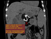
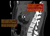

There is a large well defined encapsulated mixed density (fat, fluid and dentiform calcification) midline retroperitoneal mass displaced pancreases anteriorly and inferiorly with encasement both of celiac trunk and SMA with no gross vascular invasion, the mass exert pressure effect on distal CBD with upstream dilation of both intra and extra-hepatic bile ducts, the mass shows no significant enhancement in postcontrast study.
Both kidneys and adrenal glands look normal to suggest fatty containing angiomyolipoma or adrenal myelolipoma respectively.
Uterine congestion and distended left gonadal vein are noted due to mass effect of the mass.
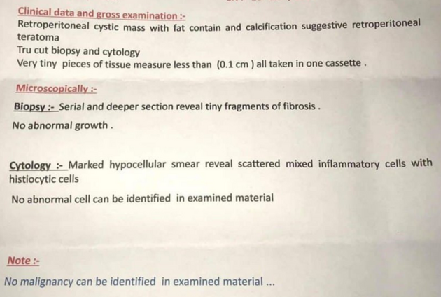
Histopathological results of true cut biopsy revealed no malignant cells.
Case Discussion
Imaging findings suggestive of benign-looking retroperitoneal teratoma which is confirmed by histopathological study. No enhancing soft tissue components to suggest liposarcoma.
Retroperitoneal teratoma is slowly growing tumor of germ cell origin. It is usually diagnosed by characteristic imaging findings of a complex mass containing a well-circumscribed fluid component of variable volume, fatty tissue and calcification in either a congealed or dentiform pattern. Histopathology is required to exclude any malignant cells.
In our case, most of patient's symptoms are related to its pressure effect of mass on nearby structure.


 Unable to process the form. Check for errors and try again.