
Presentation
Abdominal pain.
Patient Data


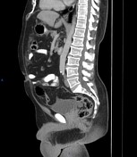
A distended appendix measures about 8 x 6 cm (appendiceal mucocele) and shows intraluminal fluid-like density with marginal calcifications. Small lesions of the same density are seen related to its tip.
Multiple cystic lesions are seen surrounding the spleen scalloping its surface, and showing variable calcifications. Similar peri hepatic lesions are also seen scalloping its surface.
Blurred anterior omentum fat with thickening (omental caking).
Free intraperitoneal collection is seen involving the paracolic gutters and pelvic recesses.
Case Discussion
Here is a case of pseudomyxoma peritonei secondary to rupture of appendiceal mucocele with suspected underlying mucinous neoplasm. Neoplasia can be of different grades: low-grade appendiceal mucinous neoplasm, high-grade appendiceal mucinous neoplasm, and mucinous adenocarcinoma.


 Unable to process the form. Check for errors and try again.