
Presentation
The patient has a red macule within the oral cavity, specifically, the left retromolar space. The lesion is non-tender, and relatively static in size on questioning.
Patient Data
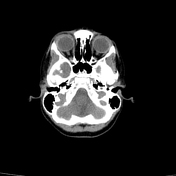

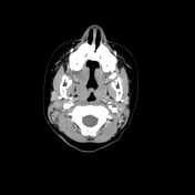
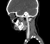
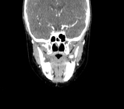

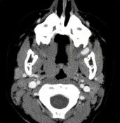
There is an intense and homogeneously enhancing, ovoid mass within the left retromolar space. The lesion is poorly appreciated in the pre-contrast study. There is no associated calcification. There are no phleboliths present. It measures 10.5 x 6.2 x 5.5 mm (AP x W x CC). There are no well-identified feeding arteries or draining veins.
There is age-appropriate adenoidal hypertrophy and palatine tonsillar prominence.
CT imaging is otherwise normal.
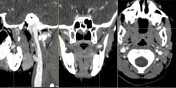
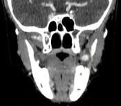
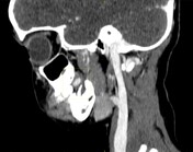
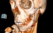
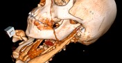
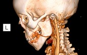
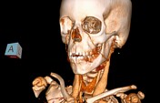
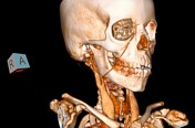
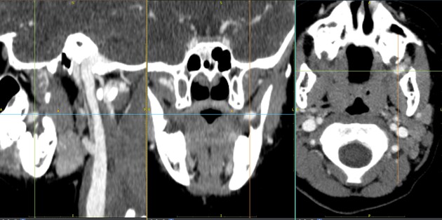
Reconstructed images demonstrate the ovoid, left retromolar, avidly enhancing lesion. The right retromolar space is normal. The lesion is expectedly identified on the 3D angiographic images, due to its intense enhancement.
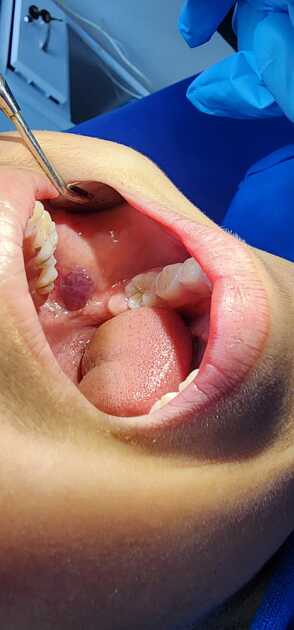
A photograph of the left retromolar, red macule at the time of clinical examination.
Photograph courtesy: Dr Z Dangor.
Case Discussion
An example of a small, retromolar space, infantile hemangioma.
Conservative management is planned due to the absence of any significant symptoms and the potential for involution.


 Unable to process the form. Check for errors and try again.