
Presentation
Three seizures at school in the setting of vomiting and not feeling well. Normal head CT.
Patient Data
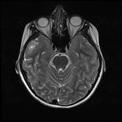


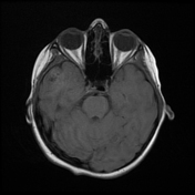

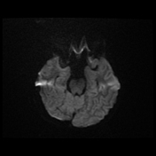

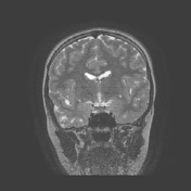
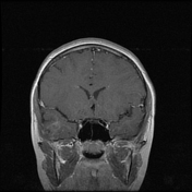
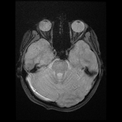
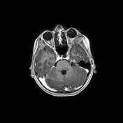
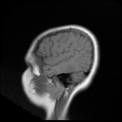
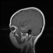
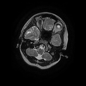
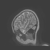

There is a mildly expansile high T2 signal lesion containing cystic areas in the right lateral temporal lobe in the middle and inferior temporal gyri. There is only mild patchy enhancement in the lesion
after contrast administration and facilitated diffusion on ADC. There is no significant mass effect.
Case Discussion
The patient underwent a craniotomy and resection of the temporal lesion.
Histology
Histopathologic analysis revealed that the mass was half parenchyma-based and half leptomeningeal-based. The major portion of the tumor consisted of fascicular glial fibers and highly pleomorphic cells typical of a pleomorphic xanthoastrocytoma, and a minor portion was made of more uniform glial cells containing microcalcifications. Mitotic activity was minimal, and no tumor necrosis or microvascular proliferation was noted.
There were small numbers of dysmorphic neurons, as those seen in epilepsy-related focal cortical dysplasia.
Stains for BRAF, CD34, EGFR, GFAP and Vimentin were positive. There was note of increased mesenchymal tissue (denoted by increased reticulin and Trichrome). The Ki-67 index is low overall. The cortical neurons and neuropil infiltrated by the tumor were highlighted by Neu-N, NF and Syn stains.
Final diagnosis: pleomorphic xanthoastrocytoma (WHO grade 2)
This is a case of an unusual pleomorphic xanthoastrocytoma, in that the majority of such tumors have vivid contrast enhancement. On imaging, the primary differential consideration should probably be a DNET or maybe a ganglioglioma.


 Unable to process the form. Check for errors and try again.