
Presentation
Seizures and memory loss.
Patient Data
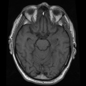

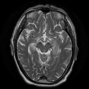

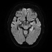
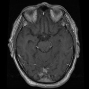

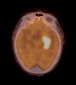
Subtle abnormal high T2 and DWI signal within the left mesial temporal lobe. No abnormal enhancement.
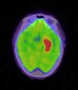
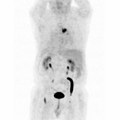
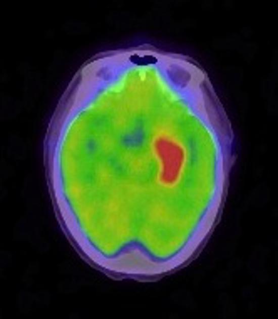
Markedly FDG avid abnormality within the left mesial temporal lobe corresponding to the MRI abnormality. An additional FDG avid focus is seen at the right lung hilum.

CT shows a right hilar / trachoebronchial soft-tissue mass corresponding to the FDG avid tissue on PET scan.
Case Discussion
The differential diagnosis for abnormal mesial temporal signal without enhancement on MRI is broad and includes infarction, HSV encephalitis, status epilepticus, low grade astrocytoma, gliomatosis cerebri and limbic encephalitis.
The marked FDG avidity on PET scan and right hilar mass however strongly favor the diagnosis of paraneoplastic limbic encephalitis. Small cell lung cancer (as was diagnosed in this case on transbronchial biopsy) is the classic cause of limbic encephalitis however many other tumors can also cause the phenomenon.


 Unable to process the form. Check for errors and try again.