
Presentation
Incidental left upper lobe mass on chest x-ray performed during an admission with a stroke.
Patient Data
Diagnostic CT performed in private sector in alternative institution without image guided biopsy facilities
Spiculated 2.4 cm nodule high in the left upper lobe suspicious for a primary lung tumor
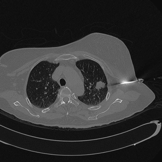
A lateral approach is chosen in this instance. It minimizes the lesion-pleural distance. The patient was unable to hold the arm above the head for an anterior approach and the scapula obstructs a posterior approach with the patient prone
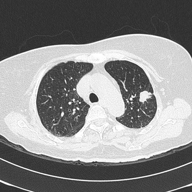
Note the breast has been taped antero-medially to reduce the distance to the mass from the lateral approach.
Markers placed in the left axilla to assist in identifying the premium slice for the biopsy approach.
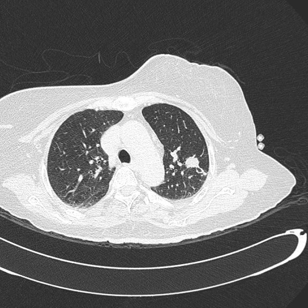
The bone window best allows identification of the needle tip. It is on course for the lesion.

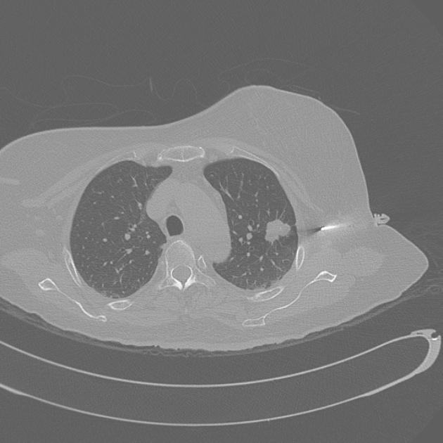
The co-axial component of the needle is 'parked' right in front of the mass to allow for the 2 cm throw of the biopsy needle.
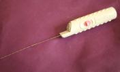
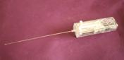

A variation of biopsy needles available for image guided biopsy procedures, such as lung biopsy.
Case Discussion
This case illustrates the step wise approach to CT guided lung biopsy. Pulmonary nodules are common and often require biopsy for histological assessment. In this case the biopsy confirmed the suspicion of lung cancer.


 Unable to process the form. Check for errors and try again.