
Presentation
Left sided parotid swelling and
Patient Data


Left sided facial lesion
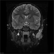
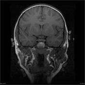
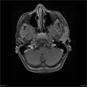
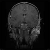
There is a large lobulated mass lesion extending from the base of skull in the peri-parotid region. It is high-T2, low-T1 and enhances. In addition, there is bilateral T2-bright white-matter abnormality.
In addition to the facial lesion, the patient has skin lesions on his back and a swollen knee. He went on to have chest imaging.
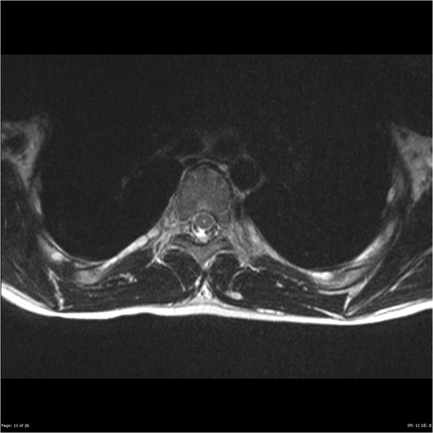
There are multiple T2-bright lesions along most of the costal nerves. Features are consistent with multiple neurofibromas.

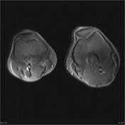

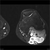
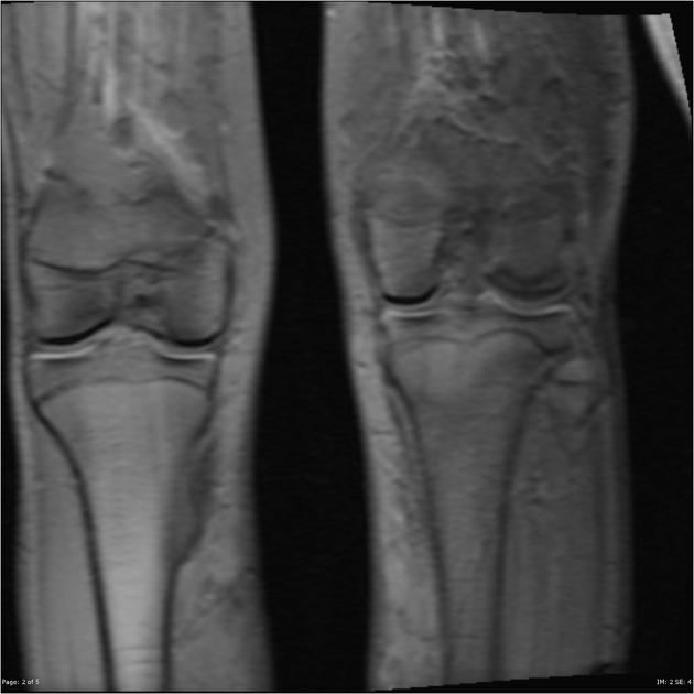
From the localizers, there is a clear size discrepancy between the two legs. On the T2 weighted imaging, the extent of the disease process is obvious. There are hugh plexiform neurofibromas extending along the femoral nerve and its branches.
Case Discussion
A case of neurofibromatosis type I with plexiform neurofibromas of the face and leg and multiple neurofibromas along the costal nerves.


 Unable to process the form. Check for errors and try again.