
Presentation
Right abdominal swelling since a week. weight loss, abdominal pain and jaundice.
Patient Data


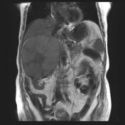

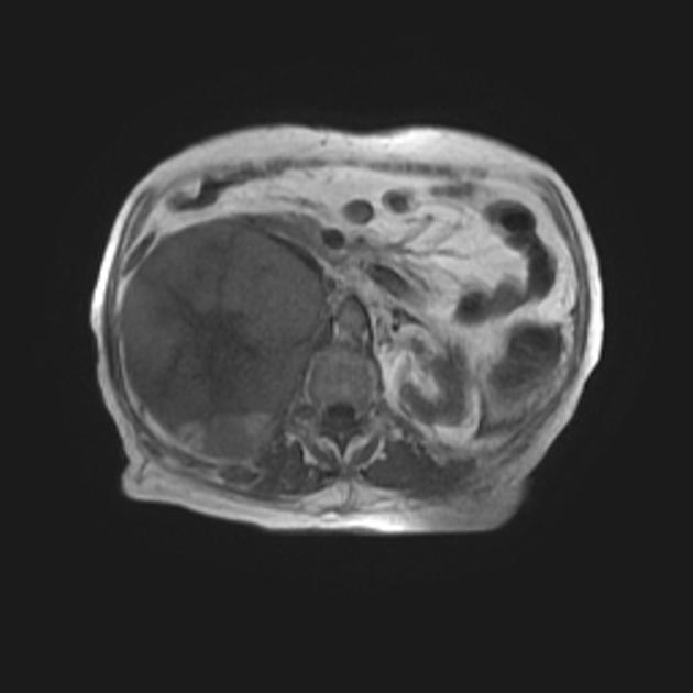
Shrunken liver with irregular nodular outline and heterogeneous parenchyma as well as hypertrophied left and caudate lobes and prominent inter-lobar fissure.
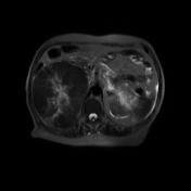
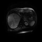
There is a intrahepatic mass lesion toards the hepatic dome juxta-caval; This is slightly ill-defined and measures approximately 6.5 X 5.5 X 5 cm. The lesion demonstrates heterogeneous; predominately hyper intense T1 and hyper intense T2 signal intensity with restricted diffusion.
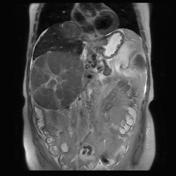
There is a much larger globular shaped lesion with central necrotic portion involving the right supra renal gland, it shows almost similar signal intensity as that of the aforementioned focal hepatic lesion. The lesion is seen compressing the right liver lobe, yet separable from it with definite plane of cleavage. It is seen however, inseparable from the right renal upper pole. The lesion is also displacing the pancreas and the bowel loops.
Enlarged porto-systemic collaterals are seen at the gastro-esophageal and peri-pancreatic regions
Enlarged spleen with no definite focal lesion.
Mild ascites.
Case Discussion
Liver cirrhosis with hepatic dome, juxta-caval malignant focal lesion; mostly hepatocellular carcinoma (HCC) with huge right adrenal heterogenous mass lesion (metastatic deposit).
Portal hypertension, splenomegaly and mild ascites.


 Unable to process the form. Check for errors and try again.