
Presentation
Known case of hemophilia presented with pain and swelling in right knee
Patient Data


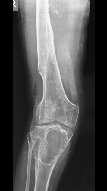
There is a well-defined, slightly expansile, osteolytic lesion seen involving proximal right tibia. The lesion is surrounded by a thin sclerotic rim and displaying thin incomplete septa-like structures.
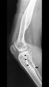
Lateral radiograph shows scalloping of the posterior margin of tibia (smaller arrows in the annotated image) and radiating trabeculae (larger arrow in the annotated image) mimicking a malignant or infectious periosteal reaction.
In a known case of hemophilia and considering the age of the patient, imaging features are most likely suggestive of an intraosseous hemophilic pseudotumor.
Case Discussion
Hemophilic pseudotumor is a rare complication occurring in 1–2% of patients with severe hemophilia.
Image differentials include:


 Unable to process the form. Check for errors and try again.