
Presentation
Chest pain and cough with mild fever.
Patient Data


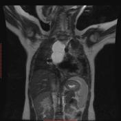
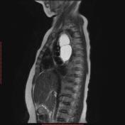
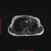
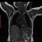
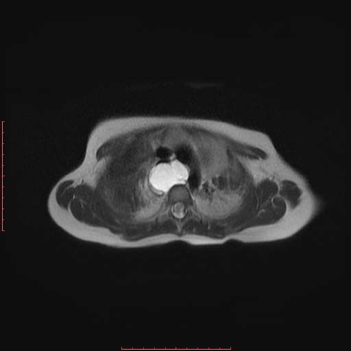
A multi-locular subcarinal posterior mediastinal cystic lesion is seen. It shows low T1 and high T2 signal intensity. The lesion measures 7 X 3.5 X 2.7 cm in its maximal cranio-caudal and axial dimensions. The lesion is seen splaying the carina and abutting the aortic arch and its main vessels. The lesion is splaying and indenting the esophagus right postero-laterally.
Bilateral lower lung lobes’ apical segments as well as left lung upper lobe apico-posterior segment and right lung lower lobe posterior segment subsegmental consolidation with air bronchogram are seen.
Normal heart and great vessels.
No hilar masses or lymph nodal enlargement.
Case Discussion
Subcarinal posterior mediastinal cystic lesion; consistent with bronchogenic cyst with bilateral pulmonary consolidations, suggestive of aspiration pneumonia.


 Unable to process the form. Check for errors and try again.